Sensor → Afferent (neurons) (towards the brain) → Brain → Efferent (axons) (away from the brain)
→ effector.
Afferent: the 5 senses (sensors): Efferent: the effector systems:
• Smell Somatomotor system:
• Sight Striated muscles (control with
• Sound conscious thought)
• Taste Visceromotor system:
• Touch: somatic (conscious world) Smooth muscles
and visceral (intestines) Glands
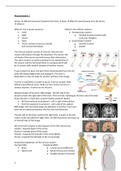
The nervous system consists of neurons. Neurons will
receive information through the dendrites. The soma is the
cell body of the neuron and will process that information.
The axon converts an action potential to the telodendria of
the neuron and the terminals there is a synapse which will
be in contact with another synapse of another neuron.
If you transect an axon, the part that is disconnected from the cell
body will always degenerate and disappear. The axon is
dependent on the cell body for protein synthesis and energy.
A nerve is essentially a bundle of axons. A nerve contains both
afferent and efferent axons. Both are from motor neurons or
sensory neurons. A nerve has no neurons.
Medical images of the brain: MRI images. The left side of the
pictures shows the right side of the brain. Point of view: Standing at the foot side of the bed.
Xrays direction is important: anterior (belly), posterior (back):
• AP (from anterior to posterior) – left is right of the patient.
• PA (from posterior to anterior) – left is left of the patient.
MRI image, front and back views are identical. A real slice, front and
back are not identical (because the slice has real thickness).
The left side of the brain controls the right body. A lesion in the left
motor cortex will affect the right limbs. On MRI that lesion will show up
on the left side of the image.
We orient with respect to the long axis of the CNS (red arrow).
Caudal = towards begin of the arrow.
Rostral = towards point of the arrow.
Ventral = towards the foreside of the arrow (belly).
Dorsal = towards the backside of the arrow (back).
Anatomical subdivision of the nervous system:
Central (CNS) Peripheral (PNS)
• Brain Cranial nerves (different)
• Spinal cord Spinal nerves (very similar)
Ganglia (clump of neurons)
Names derived from embryology: →
The neuron tube is hollow and this hollow
will develop inside the brain with cavities
and ventricles.
Learn constructions by heart.
The brain
Telencephalon:
• Two hemispheres
• Diencephalon
Brainstem:
• Mesencephalon
• Pons & Cerebellum
• Medulla obiongata
Two hemispheres are coupled by diencephalon
to the brainstem. Dissection of the brain: one
hemisphere with a half diencephalon.
The cerebellum is attached to the dorsal part
of the brainstem. The cerebellum also consists
of two hemispheres. The brainstem is
connected to the diencephalon.
Cerebral lobes are named after the part of the bone (skull).
The central sulcus separates the frontal lobe form the parietal lobe.
The lateral fissure separates the temporal lobe from parietal and frontal lobe.
Sulcus:
• Sulcus centralis cerebri: Central sulcus
o Fairly shallow; lined by neocortex only
• Sulcus calcarinus:
o Deep; lined by neocortex only Parietal
Frontal
Occipital
Fissure:
• Fissura longitudinalis cerebri:
Temporal
o Very deep; lined by neocortex, allocortex and callosal body
• Fissura lateralis cerebri:
o Deep and extensive; lined by neocortex and allocortex Lateral fissure
Always name them sulcus is the best you can do.
In front of the central sulcus is the precentral gyrus (M1: primary somatomotorcortex)) in the frontal
lobe. Behind the central sulcus is the postcentral gyrus (S1: primary
somatosensory cortex) in the parietal lobe. These are areas of the
cortex and each part of the cortex has its own specific function.
2
Voordelen van het kopen van samenvattingen bij Stuvia op een rij:
Verzekerd van kwaliteit door reviews
Stuvia-klanten hebben meer dan 700.000 samenvattingen beoordeeld. Zo weet je zeker dat je de beste documenten koopt!
Snel en makkelijk kopen
Je betaalt supersnel en eenmalig met iDeal, creditcard of Stuvia-tegoed voor de samenvatting. Zonder lidmaatschap.
Focus op de essentie
Samenvattingen worden geschreven voor en door anderen. Daarom zijn de samenvattingen altijd betrouwbaar en actueel. Zo kom je snel tot de kern!
Veelgestelde vragen
Wat krijg ik als ik dit document koop?
Je krijgt een PDF, die direct beschikbaar is na je aankoop. Het gekochte document is altijd, overal en oneindig toegankelijk via je profiel.
Tevredenheidsgarantie: hoe werkt dat?
Onze tevredenheidsgarantie zorgt ervoor dat je altijd een studiedocument vindt dat goed bij je past. Je vult een formulier in en onze klantenservice regelt de rest.
Van wie koop ik deze samenvatting?
Stuvia is een marktplaats, je koop dit document dus niet van ons, maar van verkoper Morgannd. Stuvia faciliteert de betaling aan de verkoper.
Zit ik meteen vast aan een abonnement?
Nee, je koopt alleen deze samenvatting voor €10,49. Je zit daarna nergens aan vast.