Lipoprotein Metabolism and Mouse Models of Atherosclerosis - 01/11/2021
Learning goals:
● Describe the most commonly used mouse models for studying atherosclerosis
pathology and understand how metabolic dysregulations in lipid fluxes underly
atherosclerosis pathology in these models
● Describe the similarities and differences between atherosclerosis pathology in mice
and humans
● Design experimental setups to study a specific aspect of atherosclerosis pathology or
treatment, taking into account the best mouse model to be used
● Describe statins and PCSK9 inhibitors as therapeutic strategy to treat atherosclerosis
and explain the mechanism of action of these therapies
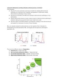
Mice are naturally resistant to atherosclerosis, as the majority of their cholesterol is
transported by the atheroprotective HDL, and largely lack the typical pro-atherogenic
lipoprotein LDL.
There are four different classes of lipoproteins:
● Chylomicrons → Triglyceride-high
● Very low density lipoprotein (VLDL) → Triglyceride-high
● Low density lipoprotein (LDL) → catabolic remnant of VLDL
● High density lipoprotein (HDL) → 50% protein, 50% lipids
,Lipoprotein classes can be separated based on size using fast-performance liquid
chromatography (FPLC). Small molecules (HDL) can enter the channels of the porous
material, whereas large molecules (VLDL / LDL) have to go around and will come through the
column first. Smaller molecules take longer. The fractured sample is analyzed by a UV
spectrophotometer for identification and concentration. Hence, this method is used to analyze
the LDL and HDL concentrations in a patient, to determine susceptibility to atherosclerosis
development.
Lipoproteins can also be separated based on density using density-gradient
ultracentrifugation. As chylomicrons and VLDL contain mostly lipids, they float on top. HDL
consists of 50% lipids and 50% proteins, hence it remains at the bottom.
Exogenous lipoprotein metabolism: cholesterol and triglycerides from our diet enter the
body through the GI tract, in which enterocytes from the intestines generate chylomicrons
from cholesterol and triglycerides. The chylomicron contains apolipoprotein B (ApoB) and
apolipoprotein E (ApoE) on its surface and enters the circulation. Next, the chylomicron is
broken down by the enzyme lipoprotein lipase (LPL) into a remnant that still contains both
ApoB and ApoE. The remnant can be taken up by the liver via the LDL receptor (LDLr),
recognizing both ApoB and ApoE, and the LRP receptor, recognizing ApoE.
The liver is a central player in the regulation of lipid metabolism. Endogenous lipoprotein
metabolism: the liver generates VLDL particles, containing triglycerides and cholesterol.
These particles have ApoB and ApoE on their surface. LPL breaks down the VLDL particles
into LDL particles, which only contain ApoB. The liver takes up the particles with LDLr and
LRP. Remaining LDL can become oxidized and taken up by arterial wall macrophages which
may become foam cells. The macrophages have scavenger receptors (SR) on their cell
surface: SR-A and SR-B1, as well as ATP-binding cassette transporter ABCA1 and ABCG1. To
prevent accumulation of these lipid-filled macrophages and atherosclerosis development, the
reverse cholesterol transport pathway comes into play.
https://www.youtube.com/watch?v=q0YiPqmsXRg
,Reverse cholesterol transport: The liver and the intestine synthesize the protein ApoA-1,
which is lipid-poor. ApoA-1 binds to the ABCA1 on the lipid-filled arterial wall macrophages,
wherafter the ApoA-1 takes up cholesterol from the macrophages and becomes a nascent
preβ HDL particle. Subsequently, lecithin cholesterol acyl transferase (LCAT) converts the
particle into a mature ɑ-HDL particle, which can take up more cholesterol via SR-B1 and
ABCG1. Mature HDL can deliver cholesterol directly to the liver via SR-B1, present on the liver
cells. In humans only, cholesteryl esters from HDL particles can be exchanged for triglycerides
in ApoB-rich particles, such as VLDL and LDL by cholesteryl ester transfer protein (CETP).
The VLDL and LDL particles can then deliver the cholesterol to the liver via the LRP or LDLr, or
go into the circulation ( → risk of atherosclerosis). This mechanism explains why humans have
more LDL than HDL, in contrast to mice.
● LDLr: recognizes ApoB and ApoE, hence takes up both VLDL particles, LDL particles,
and chylomicron remnants.
● LRP: recognizes ApoE, hence takes up VLDL particles and chylomicron remnants.
Beverly Paigen was a pioneer in atherosclerosis analysis using mice models. She put C57BL/6
mice on a high-fat diet, also known as the Paigen diet, to generate atherosclerosis in
wild-type mice. The mice developed small atherosclerotic lesions in the aortic root after 8
weeks.
Next, scientists discovered a novel mouse model for atherosclerosis: ApoE knockout mice
spontaneously developed atherosclerosis after 9-12 weeks while on a regular chow diet.
ApoE resides on VLDL particles and is an important ligand for LRP and LDLr. The VLDL
particles, however, still contain ApoB which is a ligand for LDLr. Hence, the mouse VLDL
, particles can now only be (partly) cleared by the LDLr, causing hypercholesterolemia and
atherosclerosis.
Another mouse model for atherosclerosis is using LDLr knockout mice. As these mice lack
the LDLr, uptake of LDL particles and (partly) the uptake of VLDL particles is impared.
Nonetheless, the VLDL particles can still be cleared via LRP. Hence, the change in the
lipoprotein profile is not as extreme as in ApoE knockout mice. To further enhance
atherosclerosis development, LDLr knockout mice can be put on a western-type diet.
For accelerated (6 weeks), local atherosclerotic plaque development in the carotid artery, a
silicone collar can be placed.
Differences mice & humans:
● In humans, lesions develop over decades, whilst in mouse models, lesions develop in
weeks to months.
● Female mice are more susceptible to atherosclerosis, whilst in humans men are more
susceptible than (pre-menopause) women.
● Major limitation: plaque rupture and thrombosis are very rare in mice.
● Normal intima of mice does not contain SMCs and connective tissue fibers, whilst
SMCs and connective tissue are important components of human atherosclerosis
development.
● In contrast to humans, the first branch of all major coronary arteries in mice are
protected from disease.


