Neuroimaging: Functional Magnetic Resonance Imaging
Task 1: The physicist in you
1. The patient is placed in a magnet
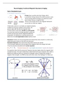
Protons have a positive electrical charge, which
is constantly moving, because the protons possess a spin.
→ This moving electrical charge is an electrical current
which always induces a magnetic field.
→ Thus, every proton has its own magnetic field and it
can be seen as a little bar magnet.
→ Normally, protons in our bodies are aligned randomly
→ When we now put a patient in the scanner, the
protons align with the external magnetic field.
→ They do this in two ways: parallel and anti-parallel.
The state that needs less energy (parallel) is preferred,
so there are few more protons aligned parallel than
antiparallel. The difference is small but present and
determines the net-magnetization vector (yellow arrow)
Precession: Protons are not just lying there but instead they move around in a certain way,
called precession (shown on the left in the blue box)
Precession frequency: how many times the protons precess per second
→ not constant, depends on the strength of the magnetic field: the stronger the faster
→ can be precisely calculated by the Larmor equation:
The equation states that the precession frequency becomes higher when the magnetic field
strength increases. The exact relationship is determined by the gyro-magnetic ratio, which is
different for different atoms (so constant for hydrogen, doesn’t change with changes in
magnetic field strength). Can fMRI be done with other atoms than hydrogen? In theory yes
,In the coordinate system, the vectors depict the protons and the z-axis the direction of the
magnetic field (the net-magnetization vector):
• Antiparallel and parallel protons can cancel each others magnetic forces out
• As there are more parallel protons, some proton’s forces aren’t cancelled out:
• All of these protons add up their forces in the direction of the external magnetic
field, so that the patient in the magnet has his own magnetic field (yellow arrow)
• This direction is longitudinal to the external field of the MR machine's magnet.
• Because it is longitudinal it cannot be measured directly (as it is in the same
direction/parallel to the external magnetic field), which is why we need
magnetization transversal to the external magnetic field
2. A radio wave is sent in
As the longitudinal magnetization cannot be measured, a radio frequency pulse is added
into the patient, to disturbs the protons, it exchanges energy with them
Resonance: Energy exchange is only possible if the protons and the RF pulse have the same
frequency (they are in resonance). This is done specifically at the Larmor frequency: the
Larmor equation gives us the needed frequency of the RF pulse
The radiowave has two effects on the protons:
1) It lifts some protons to a higher level of energy (protons pick energy up)
→ More protons flip to the energy costly anti-parallel position
→ results in decreasing the longitudinal magnetization
2) it causes the protons to precess in phase
→ this creates a new magnetization transversal to the external magnetic field
→ can now be measured: magnetic field precesses → induces an electrical current → this
current has a frequency that can be measured, this is our MR signal!
If the number of parallel protons equals the number of antiparallel protons, longitudinal
magnetization disappears, and there is only transversal magnetization (due to phase
coherence). The net magnetic vector (yellow arrow) seems to have been "tilted" 90° to the
side. The corresponding RF pulse is thus also called a 90° pulse.
,3. The radio wave is turned off
As the radio wave is turned off, the protons fall back into their original position:
Longitudinal relaxation time (T1): the time it takes for the 63% of the longitudinal
magnetization to increase and be fully back again (graph on the left of the green box)
(protons give energy back to their surroundings), 300-2000ms. It is a measure of the time
taken for spinning protons to realign with the external magnetic field.
• T1 varies with the magnetic field strength and is longer in stronger magnetic fields
because in a stronger magnetic field the protons precess faster.
• when they precess faster, they have more problems handing down their energy to a
lattice with more slowly fluctuating magnetic fields.
• fatty tissue has a shorter T1 as water because the carbon bonds at the ends of the
fatty acids have frequencies near the Larmor frequency, thus resulting in effective
energy transfer.
Transversal relaxation time (T2): the time it takes for 63% of the transversal magnetization
to decrease and fully disappear (graph on the right of the green box), 30-150ms. It is a
measure of the time taken for spinning protons to lose phase coherence among the nuclei
spinning perpendicular to the main field
• assumes homogeneity of the external magnetic field and local magnetic fields (small
imperfections)
• water molecules move around very fast → their magnetic fields fluctuate fast and
cancel each other out → no big difference in internal magnetic fields → protons stay
longer in phase → T2 is longer
• with larger molecules, there are bigger variations in the local magnetic fields:
move slower → no cancelling out → larger difference in internal magnetic fields →
larger differences in precession frequencies → protons get out of phase quicker
→ T2 is shorter
Transverse magnetization decays much faster than would be predicted by natural atomic
and molecular mechanisms: this rate is called the T2* effects:
• In an ideal homogeneous magnetic field, the transverse relaxation follows an
exponential signal decay (free-induction decay, FID), this is T2
• However, in physiological tissue the transverse relaxation is more rapid because of
local field inhomogeneities including those caused by the tissue itself. When
inhomogeneities are present, the decay constant is called T2*
• T2* can be considered an "observed" or "effective" T2, whereas the first T2 can be
considered the "natural" or "true" T2 of the tissue being imaged.
• T2* is always less than or equal to T2.
Free Induction Decay: The increasing of longitudinal and decreasing of
transversal magnetization looks like a spiraling of the net magnetic
vector. This induces an electrical current.
→ This however only applies to a 90-degree RF pulse
→ the signal decays very rapidly and requires a very fast scanner
→ T2* effects that come with the local homogeneities of the magnetic
field
, • The time of T1 and T2 differs between white and grey matter and cerebrospinal fluid. T1
and T2 take longer for watery tissue, and shorter for fatty tissue. By recording the
contrast at the moment of the maximal difference of T1 and T2 you can measure what
matter the tissue has.
• In total, T1 is longer than T2
Watch: https://www.youtube.com/watch?v=jLnuPKhKXVM
https://www.youtube.com/watch?v=chsP2mAng3o


