Synaptic plasticity chapter 8
Learning objectives: Synaptic depression
• Be able to discuss the different forms of short-term synaptic The opposite of synaptic facilitation is synaptic depression, which
plasticity causes a decline in neurotransmitter release during synaptic
• Explain how calcium shapes synaptic plasticity by affecting activity. Depression depends on the amount of neurotransmitter
neurotransmitter release that has been released. The total amount of depression is
• Describe Aplysia’s behavioural responses during habituation and proportional to the amount of transmitter that is released from
sensitisation the presynaptic terminal. This means that depression is caused by
• Explain the neuronal circuitry that facilitates gill withdrawal the depletion of a pool of synaptic vesicles that are available for
and sensitisation release. When rates of release are high, the vesicles deplete
• Know the molecular pathways that govern short- and long- rapidly and cause a lot of depression.
term sensitisation
• Have a basic understanding of the hippocampal neurocircuitry Synaptic potentiation and augmentation
from the entorhinal cortex to CA1 pyramidals These types of plasticity serve to increase the amount of
• Be able to describe the three principles of long-term synaptic transmitter released from presynaptic terminals. Both
plasticity. Namely, specificity, state dependence and associativity augmentation and potentiation enhance the ability of incoming
• Understand the contribution of AMPA and NMDA receptors and calcium to trigger the fusion of synaptic vesicles with the plasma
the downstream signalling in LTP membrane, but the two processes work over different timescales.
• Recognise and understand the differences in the downstream Augmentation works throughout a few seconds and potentiation
mechanisms between LTP and LTD acts over a timescale of seconds to minutes.
Chemical synapses are capable of undergoing plastic changes Overview
that either strengthen or weaken synaptic transmission. Synaptic
plasticity mechanisms occur on timescales ranging from
milliseconds to days, weeks, or even longer. There are two forms
of synaptic plasticity:
• Short-term plasticity (presynaptic side)
• Long-term plasticity
Short-term synaptic plasticity
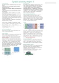
Several forms of short-term synaptic plasticity differ in time In this graph, you can see that when there is stimulation there is
courses and underlying mechanisms. facilitation, augmentation and eventually potentiation. Due to the
potentiation and the constant stimuli, the vesicle pools are
Synaptic facilitation depleted and there is depression. When the constant stimulation
Synaptic facilitation is a rapid increase in synaptic strength that stops for a few milliseconds the vesicles have time to be refilled.
occurs when two repeated action potentials invade the After a few minutes, there is a membrane potential that is still
presynaptic terminal within a few milliseconds of each other, a quite high due to the calcium residues that were present. They
paired pulse facilitation. This causes a calcium influx and the
·
call this post-tetanic potentiation (PTP).
release of a synaptic vesicle. The repeated action potentials
cause a calcium build-up which allows more synaptic vesicles to Long-term synaptic plasticity
be released. If you would chance the interval of the stimuli you This type of plasticity lasts for longer than 30 minutes and is
see that when the time between two pulses is increased, there responsible for changes in brain function that persist for weeks,
is less facilitation and there are less vesicles being released. months, or years. Because of their duration, these forms of
After 15ms there is no facilitation anymore. The target of this synaptic plasticity may be correlated with learning and memory.
calcium signal is synaptotagmin, which is a calcium-binding protein
that is found on the plasma membrane (also on synaptic vesicles Behavioural modification in Aplysia
where they function as calcium sensors and trigger The Aplysia is a sea slug that has a very simple nervous system.
neurotransmitter release). Aplysia exhibits several forms of behavioural plasticity.
• Habituation: causes the animal to become less responsive to
repeated stimuli (a form of depression)
• Sensitisation: this allows the animal to enhance a response
upon aversive stimulus (a form of facilitation)
Residual calcium
,A light touch to the siphon of the Aplysia results in the The same serotonin-induced enhancement of glutamate release
withdrawal of the animal's gill (it contracts), but habituation also mediates long-term sensitisation. However, during long-term
causes the gill contraction to become weaker during repeated sensitisation, this circuitry is affected for up to several weeks.
stimulation of the siphon. When you touch the tail and the This prolonged duration of this form of plasticity is evidently due
siphon, the gill contracts again and causes sensitisation. Even to changes in gene expression and protein synthesis.
after a single stimulus to the tail, the gill withdrawal reflex
remains enhanced for at least an hour. CREB-dependent gene expression
With repeated training (repeated tail shocks), the serotonin-
activated PKA involved in short-term sensitisation also
phosphorylates and actives the transcriptional activator CREB,
which binds to cAMP response elements (CREs) in regulatory
regions of nuclear DNA and increases the rates of transcription of
downstream genes.
This can be viewed as a simple form of short-term memory. When you would train the Aplysia with repeated stimulation over
With the repeated pairing of tail and siphon stimuli, the weeks, eventually PKA activates CREB (transcription factor) that
behaviour can be altered for days or weeks demonstrating a stimulates the synthesis of the enzyme ubiquitin hydrolase. This
simple form of long-term memory. enzyme can cleave a part of the PKA which makes the PKA auto
active which means that PKA is active and no longer needs
serotonin for the activation. CREB also stimulates another
transcriptional activator protein called C/EBP, which stimulates
the transcription of other genes that cause the addition of more
synapses and also make them bigger.
Although hundreds of neurons are involved in producing this
simple behaviour, the activities of only a few types of neurons
can account for gill withdrawal and its plasticity. These
neurons include mechanosensory neurons that innervate the
siphon, motor neurons that innervate muscles in the gill, and
interneurons that receive inputs from a variety of sensory
neurons.
Long-term potentiation at hippocampal synapses
Information goes from the perforant path from the cortex to the
granule cell in the dentate gyrus. From there the information is
sent to the CA3 pyramidal cell, and the CA3 neuron sends signals
to the CA1 pyramidal cell. These are all glutamatergic
connections. The bundles of axons between the CA3 and CA1
neurons are called the Schaffer Collaterals.
Interneuron: serotonin
Sensory neuron: glutamate
Motor neuron: Acetylcholine
When the tail is stimulated the interneuron releases serotonin,
which is an excitatory molecule. The serotonin binds to the
receptors on the sensory neuron where adenylyl cyclase is
activated and starts generating cAMP, cAMP activates protein Researchers looked into the Schaffer collaterals and stimulated
kinase A (PKA). PKA starts to phosphorylate the potassium the bundles of neurons. They saw that when they stimulate
channels on the sensory neuron that close/deactivate and pathway 1 with a few seconds of high frequency that they can
induce depolarization. Calcium channels will stay open longer enhance the synaptic transmission in the hippocampus over a
and therefore calcium will flux in and an action potential is longer time. This pattern of long-lasting synaptic activity is known
generated. as long-term potentiation (LTP).
,Specificity The specificity of the LTP can be explained by the fact that
You can see that when one pathway is stimulated, the other NMDA channels will be opened only at synaptic inputs that are
pathway shows no changes, which makes the LTP very specific. active and releasing glutamate, thereby confining LTP to these
sites even though EPSP generated at active synapses depolarises
State dependency, a coincidence detector the postsynaptic neuron. The associativity can be explained by the
When both pathways are paired and stimulated, the presynaptic fact that a weakly stimulated input releases glutamate but cannot
neuron generates an action potential and releases glutamate, but sufficiently depolarise the postsynaptic cell to relieve the
within 100 ms the postsynaptic neuron also fires an action magnesium block. If a large number of other inputs are strongly
potential. This also causes LTP of the connection. stimulated they provide the associative depolarisation necessary
to relieve the block. Meaning that the induction of LTP relies on
the activation of NMDA receptors
LTP in AMPA receptors
The expression of LTP relies on changes in the AMPA receptors.
LTP is due to the synaptotagmin-mediated insertion of AMPA
receptors into the postsynaptic membrane. The increase in AMPA
receptors increases the response of the postsynaptic cell to
Association release glutamate, yielding a strengthening of synaptic
Weak stimulation of a pathway will not by itself trigger LTP. transmission that can last for as long as LTP is maintained. LTP
However, if one pathway is weakly activated at the same time does not affect the number of postsynaptic NMDA receptors.
that the neighbouring pathway on the same cell is strongly Stimulus-induced changes in AMPA receptors can even add new
activated, then both synaptic pathways undergo LTP. The selective AMPA receptors to silent synapses so that the synapse can
enhancement of paired synaptic input is often considered a produce postsynaptic responses. Thus, the strengthening of
cellular analog for associative learning, where two stimuli are synaptic transmission during LTP arises from an increase in the
required for learning to take place. sensitivity of the postsynaptic cell to glutamate.
CaMKII and PKC
Calcium also activates signal transduction cascades that activate
calcium/calmodulin-dependent protein kinase type II (CaMKII) and
protein kinase C (PKC). CaMKII is activated by stimuli that induce
Mechanisms underlying long-term potentiation LTP and pharmacological inhibition or deletion of CaMKII prevents
This is a postsynaptic phenomenon. During LTP the NMDA LTP. CaMKII and PKC phosphorylate downstream targets that
receptors function as a coincidence detector for modulation of includes AMPA receptors.
the connection. In a normal resting potential, there is a
magnesium block on the NMDA receptor. During low-frequency
synaptic transmission glutamate is released and binds to the
AMPAR and NMDAR, but due to the magnesium block, the EPSP is
only mediated by the AMPAR. When there is high-frequency
stimulation and prolonged depolarisation and release of glutamate,
the magnesium is expelled from the channel, which causes a
calcium influx. Injection of calcium chelators blocks LTP induction
but the elevation of calcium in the postsynaptic neurons causes Long-term depression
synaptic transmission. A rise in postsynaptic calcium concentration When you stimulate a neuron for 15 minutes with low frequency,
serves as a second messenger signal that induces LTP. long-term depression is initiated. This is due to low calcium influx,
because of this phosphatases are activated instead of kinases. This
makes the AMPA receptors more mobile, they leave the spine
head, so there is a decrease in these receptors.
, The somatosensory system chapter 9
The human brain is built of neurons. The human cortex consists of Sensory receptors are activated by the energy of the stimulus
about 86 billion neurons that each have around 1000-10.000 Non-sensory neurons (brain) receive information from the axons of
synapses. Neurons communicate with each other via synaptic other neurons. Sensory neurons do not have these axons, so they
transmission via the synaptic vesicle cycle. There are two kinds of need cells that react to the stimulus (for example rods and cones
potentials, the excitatory postsynaptic potential (EPSP) which is in the retina that react to light).
regulated by the neurotransmitter glutamate that binds to the
AMPA and NMDA receptors, and the inhibitory postsynaptic
potential (IPSP) which is regulated by the neurotransmitter GABA
that binds to GABAergic neurons.
Synaptic transmission
Axons release neurotransmitters that bind to postsynaptic
receptors and determine whether the postsynaptic cell membrane Types of sensory receptors
will depolarise and generate an EPSP or hyperpolarise and • Mechanical: touch and proprioception, hearing, balance. A
generate an IPSP. EPSPs increase the excitability of the cell and physical stretch or tension on the receptor deforms the membrane
increase the probability of action potential generation. IPSPs and opens the channels.
decrease excitability and probability of action potential • Chemical: pain, itch, smell, taste. Regulated by binding to a
generation. In the neuronal network each neuron will integrate chemical receptor.
multiple EPSPs and IPSPs, the sum will determine whether the • Photoreceptors: vision, photoreceptors in the retina. There is a
cell fires an action potential. The frequency of the action change in the conformation of photosensitive proteins.
potential conveys the signal. Drugs can also bind to the receptors • Thermal: temperature
and stimulate the action of neurotransmitters. Receptor activation results in the change of postsynaptic potential
Perception of the outside world Skin mechanoreceptors
The sensory system receives and translates information into the There are four types of receptors in the skin:
language of the system: sensory receptors/axons translate the 1) Merkel cells
energy of the stimulus into electrical signals. After this the 2) Meissner corpuscle
information is transported for processing: axons transport the 3) Ruffini corpuscle
signal to the series of relay nuclei and processed information gets 4) Pacinian corpuscle
integrated: interneurons and local circuitry in nuclei process the
signal. An example of this is the receptors in the skin that can
sense a stretch when the skin is touched. This opens ion channels
and lets ions in and generates a depolarising current and an
action potential. Axons are used to transport the signal to the On the right, you can see the different responses of the different
dorsal root ganglion cells that lay close to the spinal cord and cells in the skin. The Merkel cells are the most accurate to
sent their axons to the skin. Finally, the information is determine the shape of the braille letters. The Pacinian corpuscle
transmitted to different circuits in the spinal cord and brain. reacts differently than the Merken cells and is not used to
determine the shape. These cells are mostly used when you for
example are playing an instrument.
Proprioception
Proprioception is the sense of the position of muscles and joints
> Axons that transmit touch sensory stimuli (mechanoreceptors) of the body. There are special proprioceptors, including muscle
are often encapsulated by special cells that help tune the axons spindles, Golgi tendon organs, and joint receptors that are located
to make them more sensitive to stimulation (lower thresholds for in the muscles and tendons that can sense this. Their axons are
action potential generation). Axons that lack these cells are wrapped around certain structures and when the muscle is
referred to as free nerve endings and are important in the stretched, the axons sense this and send a signal to the brain, so
sensation of pain. the brain knows where the muscles are and complex movements
can be accurately performed.
Sensory systems
1) Reception: sensory receptors translate the energy of the
stimulus into electrical signals (modality (type of the stimulus),
location, intensity, and timing)
2) Transport: axons transport the signal to the series of relay
nuclei (parallel processing, topographical representation, cross-
over, feedback connections)
3) Processing: interneurons and local circuitry in nuclei process
the signal