100% tevredenheidsgarantie Direct beschikbaar na je betaling Lees online óf als PDF Geen vaste maandelijkse kosten
logo-home
Samenvatting Fundamentals of Neuroscience, UvA €7,48
In winkelwagen

College aantekeningen

Samenvatting Fundamentals of Neuroscience, UvA

 1 keer verkocht

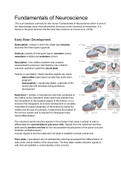
This is an extensive summary for the course Fundamentals of Neuroscience which is part of the Neurobiology minor (from Biomedical Sciences) at the University of Amsterdam. It is based on the given lectures and the book Neuroscience by Purves et al. (2018). Dit is een uitgebreide samenvatting voo...

[Meer zien]
Voorbeeld 4 van de 49  pagina's

  • 21 februari 2023
  • 49
  • 2022/2023
  • College aantekeningen
  • Tonny mulder
  • Alle colleges
book image

Titel boek:

Auteur(s):

  • Uitgave:
  • ISBN:
  • Druk:
Alle documenten voor dit vak (2)
avatar-seller
isahartman
Fundamentals of Neuroscience
This is an extensive summary for the course Fundamentals of Neuroscience which is part of
the Neurobiology minor (from Biomedical Sciences) at the University of Amsterdam. It is
based on the given lectures and the book Neuroscience by Purves et al. (2018).


Early Brain Development:
Gastrulation = phase in which the single layer blastula
becomes the three layered gastrula.

Gastrula consists of three germ layers: ectoderm (outer),
mesoderm (middle) and endoderm (inner).

Neurulation = the midline ectoderm that contains
neuroectoderm precursor cells thickens into a distinct
columnar epithelium called the neural plate.

Failures in neurulation / faulty inductive signals can cause:
- Spina bifida (open back) (so take folic acids when
pregnant)
- Anencephaly = neural tube defect, a disorder of the
neural tube that develops during embryonic
development.

Notochord = cylinder of mesodermal cells that condenses at
the midline as the mesoderm folds inward and extends from
the mid-anterior to the posterior aspect of the embryo. It is a
structure that disappears once early development is complete.
It specifies the basic topography of the embryo by being the
midline and the axis of symmetry. It determines the position of
the nervous system and is required for subsequent early
neural differentiation.

The notochord sends inductive signals to the ectoderm that cause a subset of cells to
differentiate into neuroectoderm precursor cells. Signals from the notochord and floor
plate specify position and fate for the neuroectodermal precursors of the spinal cord and
hindbrain (achterhersenen).
It sends signals to the floor plate and roof plate to establish a dorsal ventral axis,

Floor plate = specialised strip of epithelial-like cells that developed from differentiation of
cells at the ventral midline of the neural tube. The floor plate creates inductive signals to
cells that will establish a ventral identity (motor circuits).

,Roofplate = a narrow strip of neuroepithelial cells at the dorsal midline of the neural tube.
Gives rise to inductive signals that establish a dorsal identity (somatic sensory neurons,
receive incoming information and integrates them within the CNS).

The floorplate and the roofplate provide signals to the developing neural tube and all but
disappear once initial nervous system development is complete.

Steps of development:
Sperm and egg fuse → cell division → blastula → polarity in blastula (three germ layers
ectoderm, mesoderm, endoderm) → three germ layers differentiate (without this only skin
and bones develop) → neural tube formation → folding → growth.

Multipotent neural stem cells = cells within the neural tube that give rise to the entire brain,
the spinal cord and most of the peripheral nervous system. They divide to produce more
precursor cells with the capacity to produce the full range of cell classes found in mature
tissues.

Neural stem cells produce neurons, astrocytes and oligodendroglial cells.

Precursor cells farther away from the ventral midline give rise to sensory relay neurons and
related interneurons in dorsal regions of the spinal cord and hindbrain. This differentiation is
also facilitated by the roofplate.

Neural crest = the region where the edges of the folded neural plate come together. It is
formed out of a population of precursor cells at the lateral edges of the neural plate. Neural
crest cells migrate away from the neural tube through a matrix of mesenchymal cells that fill
the spaces between the neural tube, embryonic epidermis and somites. Neural crest cells
follow different pathways, along which they are exposed to different signals that influence
their specific differentiation. They give rise to neural and non-neural structures.

After the formation of the neural tube, the anterior part forms a crook (“crane handle”) by
folding and bending.

Sensory organs are formed from the placodes and dorsal ventral axis.

Placodes = embryonic structures that give rise to structures such as hair follicles, feathers
and teeth. Form eyes and ears.

Progenitor domain → develop placodes and develop eyes, ears and olfactory system.

Commissural plate ends up being the division between the two hemispheres. If the
development doesn't go right, only one big hemisphere is formed.

Proliferation in the developing brain is caused by neural precursor cells.

Symmetrical division: produces 2 precursor cells
Asymmetrical division:
- produce two different kinds of cells

, - progenitor cells (= cell that can differentiate into a specific cell type) → stages of
mitosis continue
- neuroblast = post mitotic cell, doesn’t divide again, gets influenced by inductive
signals. It is the undifferentiated precursor of the CNS.
Migration of the cell to pial surface:
- synthesis stage
- DNA is replicated
Migration back down to the neural tube:
- getting ready for division: mitosis

Grey matter = cellichamen en cel-uitlopers (dendrieten en axonen) van de neuronen. Has
layers of cells.
White matter= glia cells, astrocytes, oligodendrocytes that make myelineschede around the
axons. White because of fatty tissue around the cells.

Pial surface = surface representing the boundary between grey matter and cerebrospinal
fluid.

Pyramidal neurons (pyramidal cells) = are neurons with a pyramidal shaped cell body
(soma) and two distinct dendritic trees. The basal dendrites emerge from the base and the
apical dendrites from the apex of the pyramidal cell body.

Developmental anatomy:
The prosencephalon develops into the telencephalon and diencephalon. The
rhombencephalon becomes the metencephalon and myelencephalon. The optic vesicle
develops from the diencephalon.
(Remember the different parts by: TEL DIE MES MET MY)
- Telencephalon
- Diencephalon
- Mesencephalon
- Metencephalon
- Myelencephalon

, Hox genes = Hox (in Humans) / homeotic / homeobox genes (in drosophila). Vertebrates
have four clusters (A, B, C, D) of homologous genes with similar functions. The pattern of
Hox gene expression causes the formation of morphological features that underlie the
progressive regionalization of the developing neural tube.

The instructions for neural stem cells to make nerve cells specific to each region comes from
neighbouring cells or tissues. Interactions between adjacent germ layers are essential for
regional and cellular identity in the developing embryo.

Neural induction = apposition of the notochord to the overlying ectoderm is essential for
establishing neuroectodermal stem cells and thus the entire nervous system. Neural
induction relies on signals provided locally by adjacent cells or tissues.

The results of inductive signalling include changes in gene expression, shape and motility in
the target cells.

Dit zijn jouw voordelen als je samenvattingen koopt bij Stuvia:

Bewezen kwaliteit door reviews

Bewezen kwaliteit door reviews

Studenten hebben al meer dan 850.000 samenvattingen beoordeeld. Zo weet jij zeker dat je de beste keuze maakt!

In een paar klikken geregeld

In een paar klikken geregeld

Geen gedoe — betaal gewoon eenmalig met iDeal, creditcard of je Stuvia-tegoed en je bent klaar. Geen abonnement nodig.

Direct to-the-point

Direct to-the-point

Studenten maken samenvattingen voor studenten. Dat betekent: actuele inhoud waar jij écht wat aan hebt. Geen overbodige details!

Veelgestelde vragen

Wat krijg ik als ik dit document koop?

Je krijgt een PDF, die direct beschikbaar is na je aankoop. Het gekochte document is altijd, overal en oneindig toegankelijk via je profiel.

Tevredenheidsgarantie: hoe werkt dat?

Onze tevredenheidsgarantie zorgt ervoor dat je altijd een studiedocument vindt dat goed bij je past. Je vult een formulier in en onze klantenservice regelt de rest.

Van wie koop ik deze samenvatting?

Stuvia is een marktplaats, je koop dit document dus niet van ons, maar van verkoper isahartman. Stuvia faciliteert de betaling aan de verkoper.

Zit ik meteen vast aan een abonnement?

Nee, je koopt alleen deze samenvatting voor €7,48. Je zit daarna nergens aan vast.

Is Stuvia te vertrouwen?

4,6 sterren op Google & Trustpilot (+1000 reviews)

Afgelopen 30 dagen zijn er 68175 samenvattingen verkocht

Opgericht in 2010, al 15 jaar dé plek om samenvattingen te kopen

Begin nu gratis
€7,48  1x  verkocht
  • (0)
In winkelwagen
Toegevoegd