From Perception to Consciousness
HC 1
Riddle: Clip about Howie: can read or can’t read (difference = distance)
Retina preprocesses signals from → Rod/cones → bipolar → ganglion cells → optic nerve, optic brain
etc.
Rods and cones sensitive to different wavelengths: 496=green, 559 =yellow, rods: 400 nm
➔ Why? Rhodopsin that receives the light had different shapes for different wavelengths (may
varieties in animal kingdom)
Rhodopsin → closing of Na+ channels so that the membrane hyperpolarization → neural signal that
is sent to bipolar → ganglion cell etc.
- Hyperpolarization and closing of na+ : in the dark na+ is open, closing
Disease: Retinal color blindness: absence of a particular cone type
- Not uncommon, 8/100 males, Many tests,
We think we always see 3D (true and both false)
Why? Cones (color) almost exclusively confined to the central part of the visual field (tightly in the
fovea, widely spaced in the periphery)
Rods are mostly in the periphery
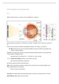
The Fovea: Cup shaped, highest density photoreceptors, mainly cones, sharpest vision, color vision
Normal Macula: bigger region around the fovea
,A lot of blood vessels over your retina, implies that whole visual field is covered with blood vessels
Fundoscopy shows the blood vessels and the fovea…
Blind spot: optic nerve/yellow spot: were all the nerves come together.
Diseases
Dry macular degeneration
Wet macular degeneration
- Symptoms: black hole (later stage) or distorted vision
- Older age, smoking, diet,
- Loss of central vision, acuity loss
- Reason: Pigment epithelium (receptors) are lost due to accumulation of toxic products
- No treatment
-
Light has to pass to the retinal network (bipolar/ganglion) to reach photoreceptors,
The photoreceptors are embedded in pigment epithelium
Why? You want a beam of light to hit only 1 photoreceptor (to have accurate/sharp vision of points
in the visual field) + Light has the tendency to scatter, so to hit other photoreceptors,
➔ retinal pigment epithelium absorbs all the light immediately, preventing scattering
Cat pigment epithelium is reflective instead of absorbent
- Better low light vision ( because same ray of light hits more photoreceptors)
- But less sharp image (due to scatter)
RGC (Retinal ganglion cell) fibers lying on top causes the blind spot the place where all retinal
ganglion cell fibers pas through the eye (optic disk) and no receptors are present
Visualizing your blind spot (close the left eye, look at your left finger, wiggle the right)
,Diseases: Glaucoma
- Increase of pressure inside the eye
- Narrow angle or open angle types /
acute, chronic
- Damage of nerve fibers on the RGC’:
optic nerve
- Loss of peripheral vision first (so you
only notice it when it already quite
bad)
- Treatment: eyedrops, surgery, (but lost
RGC’s are lost)
- Flow of ocular fluid disturbed
→increased pressure → Pushes optic
nerve a little bit out → lose optic
fibres, blood vessels get compressed
(less able to send blood)
- Normal flow ocular fluid: ciliary body
produces fluid, goes through eye, and
then out through trabecular meshwork
Processing
Why so many layers of cells?
➔ The need for data compression
From 130 million photoreceptors
To 1 million nerve fibres (axons of ganglion cells) in optic nerve
How is the retinal information compressed?
1. Contrast coding (ON and OFF center surround cells)
gradual transition vs. sharp transition of black/white: the eyes are more sensitive for sharp contrast
retina is primarily interested in change: if it doesn’t change no signal after time (stabilized vision)
The photoreceptor responds to light by hyperpolarization (closing of NA+ channels), respond to dark
by depolarization (opening of Na+ channels): a graded potential signal (No Action potential! Just
polarization or depolarization)
So amount of light is reflected in amount of hyper/depolarization.
The graded potential is transmitted by neurotransmitters based on amount of polarization
The photoreceptor signal is converted in to ON and OFF signals at the bipolar cells, using different
Glutamate receptor types at the synapse between receptor and bipolar
, A schematic of how the ON and OFF ganglion cells arise
The photoreceptors of the vertebrate retina all hyperpolarize to light, yield only grades potentials,
and utilize the neurotransmitter glutamate
- Sign conserving synapse: ON and OFF systems originate at the level of the bipolar cells,
receptor make this synapses with the OFF bipolar cells
- Sign inverting synapses: with ON bipolar cells that have a unique neurotransmitter receptor
site polarization and then depolarization??
The receptors of the Glutamate determine if the bipolar cells depolarizes or hyperpolarizes
Horizontal cells receive signals from widespread region
of receptors. They provide negative feedback on the
receptors
This gives the center-surround profile of bipolar (and
subsequently ganglion cell) receptive field
Horizontal cells add wide field (sensitive to dark around
the central field), with a peak in the central: light
(center), (or with dark the other way around)
Receptor provides + or – because of hyperpolarization or
depolarization → horizontal cells add negative feedback
so with light in the center and dark in the surround the
horizontal cells make the contrast bigger by negative
feedback (more + in the center)