100% tevredenheidsgarantie Direct beschikbaar na betaling Zowel online als in PDF Je zit nergens aan vast
logo-home
Summary Biological Psychology, everything you need to know. €7,69
In winkelwagen

Samenvatting

Summary Biological Psychology, everything you need to know.

 56 keer bekeken  5 keer verkocht

This is a summary of all the things you need to know. No excessive amounts of information. Just the things you need to know explained and made simple. Every topic is discussed. Got an 8.2

Voorbeeld 3 van de 28  pagina's

  • Ja
  • 23 maart 2023
  • 28
  • 2022/2023
  • Samenvatting
  • introduction
  • structure
  • vision
book image

Titel boek:

Auteur(s):

  • Uitgave:
  • ISBN:
  • Druk:
Alle documenten voor dit vak (1)
avatar-seller
frederikesmits
Biologische Psychologie Moeilijk
Hoorcollege 1
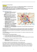
Oligodendrocytes: Form the myelin sheaths in the central nervous system. One oligodendrocytes
can apply multiple myelin sheaths to multiple axons (see figure above).
Schwann cells: Form the myelin sheaths in the peripheral nervous system. One schwann cell can
only create a myelin sheath for one axon (it wraps around it (see figure on the right

Global structure of the cell
1. Cell nucleus: Contains the DNA and
can transport mRNA with the little
pores.
a. mRNA (Messenger RNA): Parts of the
DNA that can be copied. Contains the
recipe for the production of proteins.
2. Endoplasmatic reticulum: Here the
mRNA is read to produce, store and
transport proteins.
3. Golgi apparatus: After producing the
proteins they need to be packed. This
is the post office for packing the
proteins (for example
neurotransmitters into vesicles).
4. Mitochondria: This is the power plant
of the cell that produces the ‘gasoline’
(ATP: Adenosine Tri-Phosphate).
5. Lysosomes: Does waste processing.
6. Microtubuli: This is the road system for transportation of neurotransmitters through the axon to
the terminal buttons.
Blood-brain barrier: meaning that blood can not directly enter the brain (no gaps in the capillaries) as
a protective function. The astrocytes place themselves against the blood vessels in the brain and by
doing that they can extract nutrients from the bloodstream. The astrocytes can give those nutrients to
the neurons.
Those astrocytes also keep storage of glucose (see figure). This creates an energy reserve for when
the brain is very active.
Astrocytes support: They have three functions;
● Provide structure and solidity to the brain (glia: glue).
● Isolate synaptic clefts; they isolate the context between neurons.
● Can take out nutrients from the bloodstream and give it to neurons (energy source for neurons).

Membrane potential is caused by a balance between two forces
- Diffusion: random motion
- Electrostatics: oppositely charged particles (+,-) attract each other

Cl- (chloride) is retained by the electrostatic force. It stays outside the cell
NA+ (sodium) driven inward by both diffusion and electrostatic forces. (membrane is
leaking for sodium, so it will leak into the negatively charged cell) → For this there is a
mechanism to transport the sodium that has leaked in to the outside. This is called the
sodium-potassium pump. → pump out sodium from the inside of the cell to the outside,


1

,maintaining low concentrations of sodium inside the cell. But, potassium ions (K+) are
pumped in. For every 3 sodium ions pumped out, 2 potassium ions are pumped in.

Resting potential
Na+ (sodium) → attracted by the electrical force into the negatively charged cell. Sodium
ions also pushed into the cell by the diffusion force.
K+ (potassium) → attracted into the cell by the electrical force, but are pushed out of the
cell by the diffusion force
A- (organic ions) → both electrical force and diffusion force push them out of the cell, but
the membrane does not let them. Cannot leave the cell.
Cl- (chloride) → are repealed outwards by the electrical force but pushed inward by the
diffusion force.

Action potential:
1. depolarisation → inside the cell is becoming less negative. Na+ flows inside the cell.
2. K+ (potassium) channels open, K+ flows out the cell, and will counteract the electrical effect of
the Na+ inflow.
3. Sodium channels close (refractory period), Na+ inflow is halted.
4. K+ keeps flowing out, cell inside returns to negative (repolarisation)
5. K+ channels close, Na+ channels return to their normal closed condition (can be opened again).
6. Following the massive outflow of K+, the membrane temporarily has an extra
negative charge → hyperpolarization

Passive conduction: the flow of electrical signals without regeneration(without new action potentials) .
Is much faster but the signal does decay strongly with distance. solution:
Saltatory conduction: axon is covered in myelin that prevents generation of action potentials. Action
potential is conducted passively through the myelin. New action potentials can be generated at the
Nodes of Ranvier, in between the myelin sheets. This process is faster and is energy efficient.

Hoorcollege 2
Exocytosis: When an action potential arrives, the vesicles (blaasjes) with neurotransmitters (synaptic
vesicles) release their content in the synaptic cleft. The postsynaptic cell will respond.
Depolarisation of the presynaptic membrane leads to the opening of ‘voltage-dependent’
calcium channels → Calcium will flow into the pre-synaptic terminal button, there it will
trigger the opening of the vesicles → neurotransmitters will flow into the synaptic cleft.

Fate of vesicles after neurotransmitters release:
- Kiss and Run
- Merge and recycle
- Bulk endocytosis

Neurotransmitter opens:
- When Na+ (sodium) channels are opened and Na+ flows into the cell:
depolarisation of the postsynaptic neuron (inside of the cell becomes less negative)
→ action potential → Excitatory Postsynaptic Potential (EPSP)


2

, - When Cl- (chloride) channels are opened and Cl- flows into the cell: inhibitory,
hyperpolarization of the postsynaptic neuron → Inhibitory Postsynaptic Potential
(IPSP)
Whether an inhibitory or excitatory potential happens depends on the neurotransmitter that is released.
Glutamate is for example an excitatory neurotransmitter (opens NA+ channels), where GABA is an
inhibitory neurotransmitter (opens CI- channels).

Metabotropic receptors:Ion channel opened in an indirect way. The receptor is not directly coupled
with an ion channel like ionotropic receptors. 6 steps:
1. The neurotransmitter binds to the metabotropic receptor.
2. Metabotropic receptor activates G protein.
3. The G protein releases an alpha subunit that activates an enzyme that produces a second
messenger.
4. The second messenger opens ion channels.
5. The second messengers affect the ions flow in/out the cell (EPSP of IPSP).
6. The second messengers can also influence other components of the postsynaptic
cell. E.g. by going to other parts of the cell like nucleus → change cell function → by
turning genes on/off.

There are 4 possible ion flows:
● Influx of Na+ (sodium) (leaking into the cell) causes depolarization (EPSP) → inside
less negative
● Efflux (leaking out of the cell) of K+ (potassium) causes hyperpolarization (IPSP) →
inside more negative.
● Influx of CI- (leaking into the cell) causes hyperpolarization (IPSP) → inside more
negative
● Influx of ca2+ (calcium) (flow into the cell). This can lead to 1) depolarization,
2) neurotransmitter release and 3) biochemical and structural cell changes (through enzymes:
protein catalyst).

Sodium = Na+
Potassium = K+
Calcium = Ca2+
Chloride = Cl-

Neurotransmitters are removed out of the synaptic cleft by: 1) diffusion, 2) reuptake ,
3) enzymatic degradation (afbraak door enzymen): with acetylcholine, 4) autoreception.
- autoreception: occurs when the neurotransmitter binds with the autoreceptors on the
presynaptic neuron.
- Autoreceptors: Regulate the production and release of neurotransmitters by the neuron.
Autoreceptors are generally inhibitory (they reduce the concentration of
neurotransmitters in the synaptic cleft). Autoreceptors are metabotropic; meaning that
they can:
- Reuptake Neurotransmitters: G proteins/second messengers activate reuptake
transporters
- Reduction Neurotransmitters release: G proteins/second messengers close
Ca2+ channels, reduced Ca2+ inflow results decreased opening of
neurotransmitter vesicles.


3

Voordelen van het kopen van samenvattingen bij Stuvia op een rij:

Verzekerd van kwaliteit door reviews

Verzekerd van kwaliteit door reviews

Stuvia-klanten hebben meer dan 700.000 samenvattingen beoordeeld. Zo weet je zeker dat je de beste documenten koopt!

Snel en makkelijk kopen

Snel en makkelijk kopen

Je betaalt supersnel en eenmalig met iDeal, creditcard of Stuvia-tegoed voor de samenvatting. Zonder lidmaatschap.

Focus op de essentie

Focus op de essentie

Samenvattingen worden geschreven voor en door anderen. Daarom zijn de samenvattingen altijd betrouwbaar en actueel. Zo kom je snel tot de kern!

Veelgestelde vragen

Wat krijg ik als ik dit document koop?

Je krijgt een PDF, die direct beschikbaar is na je aankoop. Het gekochte document is altijd, overal en oneindig toegankelijk via je profiel.

Tevredenheidsgarantie: hoe werkt dat?

Onze tevredenheidsgarantie zorgt ervoor dat je altijd een studiedocument vindt dat goed bij je past. Je vult een formulier in en onze klantenservice regelt de rest.

Van wie koop ik deze samenvatting?

Stuvia is een marktplaats, je koop dit document dus niet van ons, maar van verkoper frederikesmits. Stuvia faciliteert de betaling aan de verkoper.

Zit ik meteen vast aan een abonnement?

Nee, je koopt alleen deze samenvatting voor €7,69. Je zit daarna nergens aan vast.

Is Stuvia te vertrouwen?

4,6 sterren op Google & Trustpilot (+1000 reviews)

Afgelopen 30 dagen zijn er 53340 samenvattingen verkocht

Opgericht in 2010, al 14 jaar dé plek om samenvattingen te kopen

Start met verkopen
€7,69  5x  verkocht
  • (0)
In winkelwagen
Toegevoegd