1 – Cystic fibrosis
CF is the most common mono-genetic disease (autosomal
recessive) in which the airway gets blocked by a thick layer of
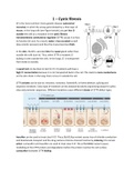
mucus. In the lung cells (see Figure beside), you get less Cl-
outside the cells as a mutation in the cystic fibrosis
transmembrane conductance regulator (CFTR) causes it to lose
its function (in-out). As a result, water is less secreted as well
(low osmotic pressure) and thus the mucus becomes thick.
In the skin, the Cl- is secreted into the sweat gland rather than
outside the cells (out-in). Thus, when CFTR is mutated, Cl-
buildup is seen outside the cells. In the lungs, Cl- is transported
from inside to outside.
A sweat test can be done to test for CF. CF patients will have a
high Cl- concentration because it is not transported back in the cell. This leads to more conductance
on the skin. Note: in the lung, there is less Cl- outside the cell.
CFTR variants can be due to: missense, nonsense, frameshift, in frame deletion, splicing and
sequence variations. Every type of mutation can be analyzed by exome sequencing except for splice
sites and promoter sequences. Different mutations cause different classes of CFTR defect types.
Ivacaftor can be used to treat class III CF. The p.Gly551Asp variant causes loss of chloride conduction
and bicarbonate transport and the drug restores chlorine channel function by inducing it to remain
active. Lumacaftor and Ivacaftor are used to treat class II CF. The p.Phe508del variant causes
misfolding of the CFTR protein and degradation before the protein reaches the cell surface.
Lumacaftor facilitates CFTR folding.
, 2 – Intellectual disabilities
Terminologies:
- Variation: any deviation from the reference genome
- Polymorphism: variation ≥1% of the allele in a population
- Mutation: variation <1% of the allele in a population
- Pathogenic: disease causing mutation or polymorphism
- CNV: deletion or duplication ≥1 kb
Types of genome variation
,Variant nomenclature
- Silent changes
when the mutation still code for the same AA → same protein (e.g. TAT > TAC codes for Tyr)
- Missense changes: there is a change in AA
- Nonsense (* = premature stop codon, no protein formed)
- Frameshift (fs)
every frameshift will lead to a premature stop codon because we only have 1 conserved ORF
- Duplication/deletion → fs → premature stop
- In frame duplication/deletion
- Splice site changes
Always bp -1 and -2 of an exon (for intronic splice site) is A and G
Always bp 1 and 2 of an intron (for exonic splice site) is G and T
- Promoter sequence variant
c. 1-333G>C
Intellectual disability
Definition:
The case where the IQ of a patient is <70 that leads to limitations in adaptive functioning and
present since <18 years.
Classification:
, ID can be caused by multifactorial causes (unlucky genes from the family) or monogenic causes
(infected brain). The mutations leading to ID can be de novo, x-linked, or autosomal recessive. ID is a
heterogeneous disorder, it can show: allelic heterogeneity or locus heterogeneity.
- Allelic heterogeneity (same gene but diff phenotype): Same/different phenotypes caused by
the same genes but with different mutations
- Locus heterogeneity: same phenotype + different genes
There are multiple ways to detect variation in the genome:
- Karyotyping → copy number variants (CNVs)
This method can be used if you think a patient has inversion. CMA is too small to see if the
chromosome is reversed.
- Chromosomal microarray (CMA) → CNVs
- Exome sequencing → CNVs, single nucleotide variants (SNVs), indels
- Genome sequencing → CNVs, SNVs, indels
3 – Chromosomal microarrays (CMAs)
Cytogenetic diagnostics for ID patients
By using genome sequencing, we can get information regarding a genetic cause of a disease in a child
and family heritage. So many genes are involved in developing major organs including the brain. So, if
there are chromosomal anomalies, most likely it can be linked to intellectual disabilities.
Karyotyping
Before 2009, we only could detect chromosomal aberrations via microscope. Metaphase
chromosomes (thickest) were used for karyotyping. Karyotyping can be used for genome-wide
testing but you can only see abnormalities when they involve millions of nucleotides (5-10 million).
Hence karyotyping has limited resolution, is difficult to automate and interpretation is subjective.
You need to be very well-trained to read the chromosomes.
If the clinical features are very specific, we can perform analysis of a specific region. If we see
symptoms of down syndrome, we immediately can test chromosome 21 (trisomy 21) to discern if it is
indeed down syndrome. For rare diseases, it would be harder to blindly guess.
FISH methodologies
Fluorescent in situ hybridization (FISH) is a method of analyzing chromosomes using labeled probes.
We get the chromosomes from the patients and heat them so they become single-stranded. Add the
probe and measure the signal and its location. This only serves as a diagnosis, and not the specific
base pair mutation. Patients can still have genetic aberration that can't be seen by microscope
(aberrant around 7%) so by using FISH, you get to observe an extra 5% aberrant.
Microarray introduction
If you don’t know where to look based on the phenotype, you can do genomic profiling using a
microarray. A microarray is a glass slide with a large quantity of (FISH) probes. Instead of hybridizing
FISH probes to DNA samples (which are labeled), we can put the DNA sample onto the microarray. A


