Molecular Principles of Development Summary Lectures 2016 – 2017
Lecture 1 Introduction and basics of Molecular Biology 10 – 11 – 2016
Overview of development, model system life cycles
Forward genetics = random mutations in sperm (radiation/use chemicals) and use the sperm to
fertilize eggs and see what happens. Then you look for interesting phenotypes.
You do not particularly look at one gene.
Reverse genetics = a gene is targeted and you mutate it and see what happens.
Major players in a genetic model system:
Gap genes
Pair rule genes
Segment polarity genes
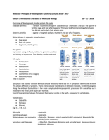
Hox genes:
Expression along A-P axis, relates to genomic position
and timing of expression. The identity can be switched.
Life cycle:
Fertilization
Cleavage
Blastulation
Gastrulation
Neurulation
(sometimes larva stages)
Metamorphosis
Time is crucial!
Syncytium is a nuclear division without cellular divisions, there is a lot of cytoplasm with nuclei in them.
This allows diffusion of proteins. There are no cellular membranes in between the cells. Gradients progress
along the embryo. Gastrulation is the more complicated morphogenetic processes, the overall lay out is
mixed and the three germ layers are formed.
Insects have an inverted axis formation, their spinal cord is in the belly, compared to vertebrates.
Vertebrates:
Zebrafish
Xenopus
Mouse
Fig. 3.3, 3.9, 3.21
Micropile – zebrafish
Germline set apart – zebrafish
Maternal axes and symmetry – Zebrafish, Xenopus: Animal-vegetal (radial symmetry), Mammals: No
polarity (point-symmetry).
Syncytium and cleavages – Zebrafish: Meroblastic divisions, yolk syncytial layer, Xenopus, mouse:
Holoblastic divisions
1
,Cell division time – Zebrafish: 15 minutes/xenopus: 25 minutes/mouse: 15-20 h
Body axes, patterning
The process of establishing positional information at the
molecular level among similar cells
Establishes body axes
Dorsal-Ventral (D-V)
Anterior-Posterior (A-P)
Medial-lateral / left-right (L-R)
Starts with polarity / symmetry-breaking
Asymmetric cell divisions
Molecular gradients
Patterning: Process which establishes differential gene expression (among otherwise similar cells) that is
directly related to position within the embryo.
Examples: A‐P, D‐V, L‐R
Patterning is not the same as differential gene expression. Patterning gives rise to differential gene
expression, but it is not the same. Patterning has to do with the molecular signals given to differentiate, it
establishes the gene expression.
Maternal factors set up the body axes. Fig. 2.9
RNA and proteins are setting up the body axes. Maternal effect genes establish the A-P and D-V axis. There
are maternally genes where zygotic genes respond to. As a result of different combinations other genes
come to expression.
Maternal products specify the A-P axis in syncytium:
Nanos protein inhibits synthesis (or translation) of Hunchback protein from maternal mRNA.
Bicoid (anterior) activates zygotic hunchback expression anteriorly, inhibits Caudal translation from
maternal mRNA. Bicoid protein inhibits synthesis of Caudal protein from maternal mRNA.
Before gastrulation, patterning of segmentation already takes place (Even-skipped, Fushi tarazu). Well
before it can be morphologically seen, the molecular models are already seen.
Germ layers, induction, fate and determination
2
, Fate ≠ Specified ≠ Determined
Fate: “we know what the cells give rise to - but the cells do not”
Specified: cells have received the instruction “cells know what they will be, but can change their mind”
Determined: “cells know what they will be and will proceed no matter what”
Fate maps tell what the progeny of cells will be.
If cells are not yet determined (mosaic), they can adapt (regulative).
Lecture 2 + 3 Origin and Specification of the germ layers in vertebrates 10 – 11 – 2016
Very early development
Maternal to zygotic transition
D-V body axis
Amphibians and fish
Maternal contribution varies greatly between fish, frogs, chicken and mammels. Mammals have an
internal egg, also known as oocyte. Chicken for example have an egg which leave the uterus, then it is
called an egg. The sperm contributes to the DNA, but in turn of RNA, protein and mitochondria they are
negligible, they are derived from the mother.
The first cell divisions you can see with the naked eye. The embryo does not grow over time; the cell
divisions start huge (xenopus).
The maternal to zygotic transition:
Maternal RNA degradation, zygotic genome activation (ZGA).
Occurs at mid-blastula stage in xenopus and zebrafish.
Early development is determined by maternal factors and after some time the factors are derived from
the embryo.
Mid-blastula transition (MBT) – a point in time when transcriptions starts
- New (embryonic) transcription
- Loss of cell cycle synchrony
- Cells become more motile
There are multiple layers of regulation. The nucleus-to-cytoplasm ratio changes dramatically during early
development. There is an increase of nuclei and the cytoplasm decreases. There will be loss of cell cycle
synchrony (happens earlier in the part with many nuclei) and a premature onset of transcription. Soon
after this stage gastrulation will happen (important cells are motile). It has to do with the balance between
nuclei and cytoplasm.
In polyspermic embryos there is more DNA in the early embryo and these start to form nuclei as well, cell
divisions happen like normal. Transcription, on the other side, is different. If there is more DNA the
transcription is starting earlier.
There are some DNA replication factors that slow cell cycle at the MBT. They are limiting for S-phase and
they force the cell cycle to slow down. If you overexpress 4 factors involved in replication this causes rapid
cell divisions and delay in transcription.
3