HOORCOLLEGE AANTEKENINGEN
NEUROLOGICAL AND PSYCHIATRIC
DISORDERS
LECTURE 1: BRAIN IMAGING
Neuroanatomy
Every lobe has a different function, which is very important in
brain imaging, because the different types of damage and
disorders that you see in the brain, affect the patient’s
differently depending on where the damage is located
Why do you want to use your imaging
- Clinic: helps with diagnosis and prognosis
o Helps with CT, MRI
- In research it helps to improve diagnostic, prediction, why things happens the way it happens
o Understand biological processes (using advanced imaging)
STRUCTURAL IMAGING
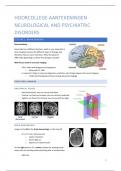
ANATOMICAL PLANES
- Axial (transverse): you can see up and down
- Coronal: cut from up to down: you can see front and back
- Sagittal: cut from front to back: you can see left and right
BRAIN HEMORRHAGE
Image on the left is the brain hemorrhage: on the top left
- It is a CT scan: 3d exray scan
o Lower resolution
o Bones light up
o Rupture of a blood vessel
On the right picture: it’s a tumor: pushes the existing tissue
away: they are pushing aside everything that is already there
- MRI scan
1
, o Better resolution
o Skull is very dark
o Based on differences in water: skull has little water so it is dark
DIFFERENT TYPES
X- ray
CT scan: When you’re in a hurry, you’ll choose CT scan→ it is much faster than MRI
MRI: When someone has long term symptoms: MRI, you can see the tissue more clear
- Identifying effects of a stroke
- Locating cyst and tumors
- Finding swelling and bleeding
- Disease related lesions
MRI (MAGNETIC RESONANCE IMAGING)
Strong magnetic field→ is on all the times→ it costs a lot to turn it of → the machine is cold
Advantages of MRI
- Non-invasive: you don’t need to put anything in the body or open it up
- Non ionizing radiation
- High soft tissue resolution and discrimination between tissue types
- Morphological (structure) information as well as functional information
Disadvantages
- Time consuming
- Contraindication for MRI: you can not scan anybody: pacemaker or some kind of surgeries, old stents
- Noise: ear protection
- Sequence need to be adapted to question → different types of scans (sequences)
HOW DOES IT WORK?
Everywhere in your body you have hydrogen atoms→ they are spinning→ randomly positioned→ when the
magnet is on they spin in the way of the magnet→ the time it takes to return that is what you measure→
different types of tissue have different amount of hydrogen atoms
- MRI uses magnetism and radiofrequency signals to acquire images
- MR images are based on density of protons
2
,MAIN TECHNIQUES
T1 weighted images: the gray matter is gray, the white matter is white: contrasts fat and water→ white matter
has a lot of fat→ you can look at different structure, you can look if the brain has damage
- Anatomy/enhancement
T2 weighted images: contrasts water and tissue→ white matter is dark, gray matter is light gray→ fluid is
white→ you can look at the pathology (it lights up when there is a disease) (T2 (tWO): water is white)
left: T1 and right: T2
T1 VS T2
T2 is sensitive for water→ white→ you can see the edema
T1: you can see the cancer what you want to see, you choose
what you want to scan
FLUID ATTENUATED INVERSION RECOVERY (FLAIR):
The scan itself is like a T2 but the water is also dark, it is easier to see the
damaged area
- T2-weighted MRI scan
- Inversion recovery: CSF is suppressed
T1 vs FLAIR
- T1: neuroanatomical changes
- FLAIR: changes related to water
3
,DIR (DOUBLE INVERSION RECOVERY)
- T2 + FLAIR
- Suppress both CSF and white matter signal
o Lesions/plaques in white matter or between gray/white matter
SUMMARY
A: T1 scan: fat/water, edema
B: T2: tissue/water, loss of membranes
C: FLAIR: T2 with CSF suppression, improves contrast
D: DIR: suppress CSF and white matter, lesions between tissue types
The stronger the magnetic field, the higher the revolution
MRA (MAGNETIC RESONANCE ANGIOGRAPHY)
Abnormalities in blood vessels: you can look where you need to perform surgery
Ischemic (less blood supply) stroke: comparing modalities
- MRA: less signal, because there is less blood supply→ on the left (right picture)
BRAIN IMAGING IN RESEARCH
Qualitative
- Standard clinical practice
- Look for pathology (not measuring, but looking at pressure, herniation, tumors etc.)
Quantitative
- Numbers as output
- Understand biological mechanisms
- Compare patient groups to healthy controls
4
,MRI: DIFFUSION TENSOR IMAGING (DTI)
- Why white matter matters?
o Alzheimer, Parkinson, Multiple sclerosis,
Schizophrenia
A: T2 scan: water is white
C: there is differences in the wire of the brain, you can predict of what
kind of symptoms you’d expect
DTI: measures how water molecules move through axons (white
matter fibre tract) in tissues: assessing white matter integrity→ water
can not move trough the walls of the axon, but it can move trough the
axon, but when the myelin is damaged, then the water can move more easily through the walls
- You can see the connections between specific brain
regions, this is used in research
- Measure the directionality in water flow
- Better flow of water means myelin present
- If the water flows in all directions, there must be
no/less myelin
- If the water flows in one direction, there is more
myelin
FOUR MAIN DIFFUSION OUTCOME PARAMETERS FOR MICROSTRUCTURAL INTEGRITY
1. Fractional anisotropy (FA): you use that to measure changes in the brain: directionality of the water
diffusion
2. Mean diffusivity (MD): the average diffusivity
3. Axonal diffusivity (AD): diffusivity along the axon
4. Radial diffusivity (RD) diffusivity peripendicular to the axon
Colors mean directions, you can look at differences in the wire of the brain
Anisotropic: characteristics depend on the directions
Isotropic: characteristics don’t depend on the directions
5
,STRUCTURAL NETWORKS
- Structural connectivity
o Quantify the number of axons between two
brain regions
▪ You can see that in patients with MS:
you can see 20.000 connections but in
healthy persons there are 40.000 for
example
FUNCTIONAL IMAGING
PET (POSITRON EMISSION TOMOGRAPHY)
- Radiotracers
- We can visualize and measure metabolic processes
- Different tracers for different purposes: 18F (fluoride), 11C (carbon), 15O (oxygen)
You give an injection with a radioactive isotope + drug→ administered into artery so it can go to the brain→
Gamma ray emission → detection
(PET- CT en PET-MRI)
- Second picture: PET scan over an MRI scan→ GLIOMA has high metabolism and uses much energy:
isotope on an glucose molecule
- Meningioma: there is some abnormality, but with a PET you can see where the tumor is and where it
is
- Lymphoma: cancerous cells: grab the energy from surrounding tissue, so there there is less energy and
metabolism
First picture: healthy picture: very clear blobs: striaium
Second picture: dopaminergic abnormality: Parkinson or lewie body dementia
6
,Second picture: hypometabolism: less metabolism: memory function→ alzheimers
Second picture: ischemic stroke: less blood supply→ tissue dead
Left: more beta amyloid uptake: with Alzheimer’s , right: healthy old person
MRS (MAGNETIC RESONANCE SPECTROSCOPY)
- Special type of MRI
- Generate a spectrum instead of an image
- Measure metabolite concentration in brain
o Choline, creatine, GABA, Glutamate, Glutathione
You get a spectrum, each peak stands for a different type of metabolite →
you can look at specific places at the brain
No ionizing radiation → it depends on what you look at if you choose PET or
MRS
FMRI (FUNCTIONAL MRI)
In your blood you carry oxygen→ hemoglobine→ it has different metabolism when it has oxygen or not →
neurons need energy: oxygen is needed→ increase in oxinated blood → as soon as you have electromagnetic
activity→ cells need more oxygen to compensate
- Hemoglobin is diamagnetic when oxygenated and
paramagnetic when deoxygenated → local MR
signal is dependent on oxygenation of blood
- fMRI measures the hemodynamic response of
neuronal activity
- BOLD (blood-oxygen-level depended contrast)
response: is a lot slower than the neural activity in
the brain
7
, TASK BASED FMRI- BLOCK DESIGN
Task based fMRI: people are doing something during the fMRI scan
Controlled block to account for al the brain activity: you really focus on the cat or dog component→ you also
show pictures of animals in general so you can substract that in the controlled block design
- Block design powerful in detecting activated voxels
(volume element, 3d pixel)
- Weak ability to determine the time course of the
response (summation of hemodynamic responses in
time)
Example: press the right button when you see a tropical landscape and left when it is not, sometimes there is
an arrow on it → it is an image, it is an landscape and you have the buttons, so you substract that so you only
have the choice of the arrow
- Encoding: 50 landscape images and 20 arrow images
- Retrieval: 100 landscape images and 20 arrow images
DISADVANTAGES FMRI
- Indirect (based on BOLD)
- Slow
- Are there other techniques?
MEG (MAGNETOENCEPHALOGRAPHY)
- MEG measures dendritic activity by measuring magnetic fields
- EEG measures electric fields in axons AND dendrites
Every electric field has also magnetic field
WHY IS MEG BETTER?
- Magnetic fields are not influenced by the scalp/skull
o Hair gel, sweating, etc
o But still influenced by metal
o Hair dye, fillings, braces
- Takes a lot less time
- Needs no reference channel
- i.e. better signal and standard setup
WHAT DOES MEG MEASURE?
Pyramidal cells
- Many in the same orientation in the cortex
Dendrites
- Fire longer
- More time to measure
8


