Developing Nervous System
Neurodevelopment → the brains development of neurological
pathways that influence performance of functioning.
Brain growth starts at week 5.
Ectoderm → develops into nervous system.
- Dorsal-most ectoderm → neural plate → rolls up forming
the neural tube (Neurulation).
- Neurulation → neural plate becomes neural tube. Neural
epithelium invaginates, dives into embryo, pinches off from
surrounding ectoderm and forms separate tissue.
- Neural crest cells from ectoderm detach and migrate to
form major part of PNS.
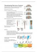
Speed of development is not uniform, gives rise to three vesicles.
- Forebrain (prosencephalon) →
tel- & diencephalon
- Midbrain (mesencephalon) →
mesencephalon
- Hindbrain (rhombencephalon) →
met- & myelencephalon
Neural canal forms ventricular system,
center of neural tube.
Mesoderm:
- Notochord → induces thickening
of ectodermal cells to form neural plate.
- Somitogenesis → development of presomitic mesoderm (PSM) lying on either side of central
notochord, divides into spherical epithelial somites, which formed out of paraxial mesoderm.
Develops anterior-posterior (AP).
Embryonic stage → neuron development
and proliferation.
Fetal stage → neuron migration and
branching, pruning, synapse reorganization,
and myelination.
-1-
,Neurogenesis
Neuronal structure in three stages:
- Cell proliferation
- Cell migration
- Cell differentiation
The brain develops from the walls of
the five fluid-filled vesicles.
- First cells that migrate to
cortical plate form the subplate.
- Neural precursors cells for layer
VI migrate collect in cortical
plate.
- Process repeats until all layers formed.
1 → cell extends a process
2 → cell’s DNA is copied at pial surface
3 → Two complete copies of DNA
4 → cell retracts arm from pia surface
5 → cell divides in two
Symmetrical cell division → both daughter cells
remain in ventricular zone.
Asymmetrical cell division → daughter cell
farthest away from ventricle ceases further division and migrates up, due to the effect of Numb.
Radial glial cells → progenitor (stem-like) cells that generate both neurons and glial cells. Divisions:
- Symmetrical division → 2 progenitor cells (expand population of proliferative progenitor cells)
- Early asymmetrical division → promotes increase neuron population
- Late asymmetrical division → promotes glia production
Daughter cells migrate along radial glial cells to the cortex where they become neurons.
Notch regulates neuronal development due to Notch. After birth Notch increases, which inhibits
differentiation of neurons and increases differentiation into astrocytes and oligodendrocytes (due
Olig1 and Olig2 expression).
Order off cell differentiation in cerebral cortex is regulated by notch signaling:
1. Neuronal differentiation (primarily pre-natal)
2. Astrocyte differentiation (peaks around birth)
3. Oligodendrocyte differentiation
-2-
,Neuronal cell differentiation
The growth cone, which are sheets of actin and microtubules, identifies the appropriate path for
neurite elongation. Neurons extend their axons to target cells, which secrete low levels of
neurotrophic factors. The neurotrophic factor binds to receptors (mostly tyrosine kinase (Trk)),
causing dimerization, and is transported to the cell body, where it promotes neuronal survival.
Neurons that fail to receive adequate
amounts of neurotrophic factor die
through apoptosis.
Nerve growth factor (NGF), Brain-
derived neurotrophic factor (BDNF),
Neurotrophin-3, and Neurotrophin-4
are factors promoting neuronal
survival.
Growth cone connects target → synapse formed.
1. Dendritic filopodium contacts axon.
2. Contact leads to recruitment of synaptic vesicles and
active zone proteins to the presynaptic membrane.
3. Neurotransmitter receptors accumulate post-
synaptically.
Formation neuromuscular synapse (PNS): postsynaptic
membrane, motor end-plate, contains junctional folds with
numerous neurotransmitter receptors.
(1) Filopodium contacts adhesive cue and contracts, pulling
growth cone forward. (2) Microtubules form central core
advance. (3) Actin polymerization pushes filopodium forward.
Differences and
similarities of PNS and CNS
Similarities Differences
Structure CNS synapses have no
basal lamina.
Clustering CNS synapses have no
neurotransmitter junctional folds but
receptors dendritic spines
Bi-directional signaling CNS synapses use
glutamate
Synaptic vesicle have PNS synapses use ACh
similar components
Synapse elimination Different
during development. neurotransmitter
receptors.
-3-
, Semaphorin 3A → a chemo-repulsive agent against axonal growth. It inhibits axonal growth, or
simulates the growth of apical dendrites. It prevents axons from growing towards the marginal
zones. Also functions as inhibitor of angiogenesis. Repels the growing axon and attracts the growing
apical dendrite.
Synaptic plasticity
The ability of synapses to strengthen or weaken
over time.
• Activation of protein kinases enhances
current through AMPA receptors
• Retrograde messengers that activate
protein kinases in presynaptic terminal
enhance subsequent transmitter release.
Events in LTP induction: (1) Glutamate binds to
NMDA and AMPA receptor, (2) NA+ enters via
AMPA receptors, depolarizing post-synapse,
relieving Mg2+ block for NMDA receptor, (3)
Ca2+ can flow in via NMDA receptor.
Events in early phase LTP: (1) CaMKII results in
more AMPA receptors inserted into the
membrane, (2) NOS produces NO, which can
diffuse back to pre-synaptic terminal stimulating
guanylate cyclase to produce cGMP, which
activates protein kinase G, which has a positive
effect on voltage-gated ion channels needed for glutamate release.
Events in late phase LTP: (1) Ca2+ activates adenylate cyclase, which produces cAMP, which
stimulates PKA, which phosphorylates CREB in nucleus, resulting in more AMPA receptors being
transcribed. (2) NOS produces NO, stimulates guanylate cyclase, stimulate cGMP, which activate PKG,
which phosphorylates CREB, resulting in more AMPA receptors being transcribed.
Long-term depression → activity-dependent reduction in neuronal synapses lasting hours or longer
following a long patterned stimulus. Selectively weaken specific synapses in order to make
constructure use of synaptic strengthening by LTP.
Synapses
Chemical synapse → electrical activity is converted into the release of a neurotransmitter which can
bind to the receptor of the postsynaptic neuron.
Electrical synapse → pre- and postsynaptic membranes are connected via gap junctions capable of
passing an electric current. Gap junctions are made out of 2 connexons, which are made out of 6
connexins each.
Peripheral synapse (neuromuscular junction) → synapses in PNS between neurons and muscles.
-4-