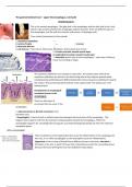
The gastrointestinal tract – upper GI (oesophagus, stomach)
OESOPHGAGUS
This is the normal oesophagus. The light pink is the oesophagus and the dark pink is the z-link,
which is the junction between the oesophagus and the stomach. There are different layers in
the oesophagus, just like with the stomach and column. Oesophagus wall:
From inside (luminal part) to the outside:
1. squamous epithelium
2. Lamina Propria = mucosa
3. Muscular Mucosa
4. sub mucosa – loose tissue, fatty tissue, fibroblasts, blood vessels and nerves.
5. Circular muscular smooth muscle layer
6. longitudinal muscular smooth muscle layer
7. Adventitia (outer layer of the oesophagus) – outer layer of fibrous
tissue surrounding an organ.
The squamous epithelium are stacked on each other. The mitotic (stem cells) of the
squamous epithelium are found on the basal lining where they migrate upwards while
simultaneously proliferating and differentiating into mature squamous epithelium towards
the surface. They eventually fall off and the circle repeats itself. The yellow part is the
lamina propria.
Development of oesophageal
carcinoma (cancer in the
oesophagus):
There are two types of
carcinoma that can occur in the
oesophagus:
1. adenocarcinoma (= Barrett carcinoma) most
prevalent one).
- Oesophagitis; it starts of with an inflammation that damages the tissue layer of the oesophagus. This
happens due to gastric acid from the stomach coming upwards towards the oesophagus. When this
oesophagitis happens for a prolonged time (so gastric acid keeps flowing backwards) we enter the intestinal
metaplasia phase.
These small dots are the lymphocytes that cause this inflammation in the oesophagus (in
this case, it is a reflex oesophagitis so the neutrophils cause this inflammation).
Hyperemia is the increase of blood flow to different tissues in the body. ulceration =
formation of an ulcer (= patch of tissue that is discontinuous with the surrounding tissue
because the tissue withing the ulcer has died/been swept away).
, during metaplasia, the normal squamous epithelium (left) differentiates into cylindrical
epithelium (right). A characteristic of cylindrical epithelium is that is forms glands (the
circular structures in the picture, also called goblet cells). These glands can produce
mucus for instance and are not neoplastic (something related to abnormal growth of
tissue). They do not have any molecular changes in their genome, so they are normal
cells but in the wrong place. Thus, metaplasia is not neoplastic; the cylindrical cells are
normal cells but in this case, they are in the wrong place (they should be located in the
intestines hence it is called intestinal metaplasia).
The little white bubbles in the gland cells are the ones that are responsible
for producing mucus (cylindrical epithelium). This usually does not happen
with normal squamous epithelium because they do not have these gland
cells which the cylindrical cells do have.
- Intestinal metaplasia: The squamous epithelial cells starts to
differentiate into glandular cells (cylindrical epithelium). Because these
glandular/cylindrical epithelial cells are primarily found in the intestines, it
is called intestinal metaplasia. The cells transform in the lining of the
upper digestive tract. Metaplasia = cells start to differentiate into a
different direction than they should supposed to follow. These cylindrical
cells in the oesophagus are not prepared for the acidic environment from
the stomach so in some cases (in some patients) it can progress into
dysplasia.
- Dysplasia: is an abnormality of development of the squamous epithelium
cells (microscopic scale) or organs (macroscopic scale) and the abnormal
histology/anatomical structure resulting from such growth. Dysplasia is
quite rare, it does not occur often. You can still see the gland cells (goblet
cells; white bubbles) but you also see large irregular shaped nuclei (purple
structures near the goblet cells) → right side of the picture. When it
progresses to a worse/higher state of dysplasia, we look at the left side of
the picture in which you see many irregular shaped nuclei and you don’t
recognize the goblet cells anymore; they have lost their differentiation.
The nuclei however are big, irregular and hyperchromatic (dark
stained). This is a state in which genomic changes have occurred; these
can be mutations or chromosomal abnormalities. The difference
between dysplasia and adenocarcinoma is that dysplasia is NOT
INVASIVE, but they do already have molecular changes in their DNA;
dysplasia is still located in the place where it should be located.
- adenocarcinoma of the oesophagus (=Barret carcinoma) = CANCER.
it is INVASIVE (you can see the tumor on the left picture).
,The white bubbles are the gland cells (globular cells). These are invading the deeper layers of the oesophagus.
So once it is invading, you can call it adenocarcinoma. . However, when they are still at the top near the
epithelial cells, then we call it dysplasia.
the left picture is the normal state; you see a clear basal lining where the
stem cells are located of the squamous epithelium and that migrate
upwards while proliferating and differentiating into normal squamous
cells. The right picture, is dysplasia. Here, the basal lining is not straight
(there is a lesion of the precursor cell lining). Also, the cells have larger
nuclei, are hypochromatin (dark stained) and are irregularly shaped; some
are large and some are small. You know that this is a dysplasia because
you can still see a clear line, indicating that there is no infiltration into the
deeper cell layers of the oesophagus.
2. Squamous cell carcinoma.
The microscopic picture in the middle, shows on the upper right part,
still a little bit or normal squamous epithelium but in the middle
there is a huge gap in which the tumor is infiltrating the deeper layers
of the oesophagus. The dark pink areas are the muscular layers of the
oesophagus and the purple-ish structures are the squamous
carcinoma that are infiltrating into the deeper parts of the
oesophagus.
Adenocarcinoma is derived from cylindrical epithelium that can occur in the stomach because there is a
precursor lesion that exists of cylindrical cells. With the squamous cell carcinoma, you have the dysplasia state
of just the squamous epithelium.
STOMACH
The stomach has a structure with folds; the function of these folds is to let the stomach extend when food
enters the stomach. The stomach is lined with cylindrical epithelium which consists of different layers from
inside → outside: gastric mucosa (mucous membrane layer of the stomach).
1. Foveolar layer – also called gastric pits.
2. Glandular layer – these harbour different cells
3. Smooth muscle layer
We know that there is also a lamina propria just below the
epithelium in the oesophagus, but in the stomach and the
bowel, the lamina propria is located between these glands
(glandular layer).
We distinguish the stomach into the corpus (upper part of the
stomach) and the antrum (lower part of the stomach). They
both contain a foveolar layer, glandular layer and smooth
muscle layer. However, the antrum does NOT have the
parietal cells (in the glandular layer. So only the corpus has
the parietal cells.
Instead, the antrum contains hormone-producing cells (e.g. g-cells that produce gastrin;
gastrin stimulates the parietal cells and the corpus to produce acid) in the glandular layer.
, types of cells that can be found in the stomach. In the foveolar layer, mucous
cells are found that produce mucus in order to protect the cells from the acidic
environment inside the stomach. In the glandular layer, we have the many
different cells of which an important one are the parietal cells that form acid and
intrinsic factor.
Inflammation of the gastric mucosa (= gastritis)
- Acute gastritis
a. Helicobacter. pylori bacterium – this bacterium does not give
any complains but on the long term, it can induce myoplasia.
b. alcohol
c. NSAID
- Auto-immune gastritis – inflammation directed towards our own cells.
a. antibodies against parietal cells located in the glandular layer can be produced
and in some cases, these antibodies can also be directed to the intrinsic factors. These
parietal cells in the end get destroyed, meaning that the stomach cannot produce acid and
intrinsic factors anymore.
These parietal cells can differentiate to a certain extent, so when the stomach cannot keep it
up anymore, these cells start to differentiate into the intestinal epithelium cells (intestinal
metaplasia).
In these patients, you don’t see parietal cells anymore, but mucous producing cells. These
patients develop anemia (= lack of red blood cells/haemoglobin). The function of the intrinsic
factor (which becomes destroyed by the antibodies) is important for the uptake of vitamin
B12 which is essential for the production of red blood cells. No parietal cells → no intrinsic
factor produced → no vitamin B12 uptake → no/decreased production of red blood cells
(=ANEMIA).
The auto-immune gastritis also increases the risk for developing adenocarcinoma
(oesophagus cancer). reason: they have this precursor lesion (intestinal metaplasia).
The auto-immune gastritis also increases the risk for developing neuro-endocrine tumors.
Development of gastric adenocarcinoma:
Majority of patients with gastric adenocarcinoma do not have an auto-immune
disease, but have this adenocarcinoma often induced by the H. pylori bacteria. In
the oesophagus, we have two types of carcinoma’s (adenocarcinoma and
squamous epithelium carcinoma). The stomach only has adenocarcinoma. There
are 2 types of gastric adenocarcinoma’s:
1. intestinal type adenocarcinoma – the
intestinal metaplasia is the precursor lesion that
progresses to the adenocarcinoma. The right
side of the purple picture shows the normal state
with the 3 layers of gastric mucosa. The left side
shows the adenocarcinoma in these layers; the
cells invade the different types of layers →
invasive, thus adenocarcinoma. When the abnormalities are only present at the top of the
mucosa, then we say that it is dysplasia.


