The eye is one of the most sensitive organ in an organism. As fixed in the eye socket, it has the bulged part protruding externally consisting of the cornea, iris, choroid
CONTE
a) Sensory recptos
b) Skin as a sen organ
c) The organ of sight, Struce of the ey
d) Functios of the ey (Image formatin and acomdtin)
e) Defcts of the ey and their coretin
SENORY RECPTOS
Al livng organism respond to changes in their enviromt (stimul). Thes changes can be mechanil, elctromagni, chemial or thermal. Thoug most cels in the bodies of organism are senitv to stimul, certain cels specialz in detcing a particul type of stimul, thes are caled sen cels or senory recptos whic are quite many in human bodies, monitrg the interal enviromt.
Sensory recptos senitv to mechanil changes are caled mechanorpts likews thermocps, chemorpts and photrecs are senitv to heat, chemial and light respctivly.
The senory recptos change the detc stimul into elctria impulse whic when recivd by the brain are transled into pictures, sound, smel, tase senatio. Struces contaig senory recptos are refd to as sen organs
A sen organ is defin as a group of specialzd cels or tisue or whic are able to reciv, perciv or detc stimul and transmi the informat to the central nervous sytem. Ther are five types of sen organs in mals; thes include
a. Skin detcing touch, pain, presu and heat or cold.
b. Eye detcing light (sen of sight)
c. Ear detcing sound (sen of hearing and balncig)
d. Nose detcing smel
e. Tongue detcing tase
THE ORGAN OF SIGHT (EY)
The ey is the organ of sight, spherical in shape and protecd by ocular or optical struce like ey socket, eylids, eylash, tear or lacrim glands and conjutiva.
a. The ey socket house the eys.
b. Eye lids (uper and lower) protec the ey from foreign particles or mechanil injury.
c. Tear or lacrim glands, at the meting point of the eylids secrt a salty fluid caled tear whic washe dust and destroy bacteri using its chemial subtance caled lysozme.
d. Eye lashe are rows of hairs on the eylid whic protec the ey bal from dust, excsiv light and shield the ey aginst sweat and water.
e. Conjuctiva is a thin transpe mebran ling the insde of the eylids and protecivly covers the cornea. The conjutiva gets inflamed during infecto (conjutivs).
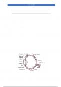
STRUCE OF THE EY
, DON BARRY
The wal of the ey bal consit of thre layers namely (from outside inwards): sclera, chorid and retina
a. THE SCLERA: -the outerms white layer whic gives shape to the ey and protecs the iner part of the ey. The scleroti layer bulges out in front of the ey to form the transpe cornea. The cornea admits light into the ey, brings the light to focus on the retina and protecs the ey extrnaly.
b. THE CHORID LAYER: – This is higly vasculrized and pigmentd (black). This layer provides fod and oxygen to the cels in the ey. The black pigment helps to absor light rays and prevnts light reflction. It consit of the cilary muscle, irs, puils, supenory ligaments and the lens.
c. IRS: – a band of muscle fibres tha contras and relaxs to alter the size of the puils therby contrlig the amount of light pasing throug the ey.
d. PUIL: – This is the openig betwn the uper and lower Iris and it contrls the amount of light whic entrs the ey. Bright light makes the puil smal while dim light makes it big.
e. CILARY MUSCLE: – It consit of cirula and radil muscle whic contras and relaxs to alter the focal length of the lens to focus near and distan objects.
f. SUPENORY LIGAMENTS: – Hold the lens in place
g. LENS: – This is a transpe biconvex elastic struce held in positn by supenory ligament. It helps to refact light rays entrig the ey. It also makes fine adjustmen to focus the image of an object on the retina.
h. THE RETINA: – This is the part of the ey senitv to light. It is also vasculrized, pigmentd and elastic. Light rays come to focus on the retina. Images formed on the retina are alwys real, invertd, and smaler than the real object. Two types of senory cels (phot recptos) found in retina are cones and rods.
a. Cones are cels in the retina, whic are senitv to colured vison and hig light intes. heyT contai a photcemial subtance caled iodpsn whic is not easily bleachd by hig light intes.
b. Rods are more than the cones. They are senitv to colures vison and low light intes. A purle pigment – protein complex made from vitamn A caled rhodpsin is found on the surface of rods. Rhodpsin is easily bleachd when light fals on it.
i. YELOW SPOT (Fovea centralis): – This is the most senitv part of the retina from wher the fulest visual informat is sent to the brain. It is the point wher image is focused.
j. BLIND SPOT: – This is the point wher the cels are not senitv to light i.e no cones or rods her. The optic nerv goes out of the ey to the brain from the blind spot.
k. OPTIC NERVS: This nerv transmi senory impulse to and from the brain.
l. AQUEOS HUMOR: – This is the transpe watery liqud whic fils the space betwn the cornea and the lens. It is made up of solutin of protein, sugar, salt and water. This liqud refacts light rays ont the retina and helps to maint the spherical shape of the ey.
m. VITREOUS HUMOR: – This is wider, transpe, jely-ik liqud whic fils the space betwn the lens and the retina. It is also a mixture of protein, sugar, salt and water. It caries out the same functio as the aqueos humor.
FUNCTIOS OF THE EY
The ey perfoms two major roles; Image formatin and acomdtin.
1. Image Formatin:-
Light rays from any object pas entr the ey throug cornea, pas throug the aqueos humor, lens and vitreous humor to the retina. Thes struce are al transpe and contribue to the refaction (bendig) of the light rays thus enablig the rays to converg on the retina. The image of the object (real, invertd and smaler) is then formed on the re tina.
Voordelen van het kopen van samenvattingen bij Stuvia op een rij:
Verzekerd van kwaliteit door reviews
Stuvia-klanten hebben meer dan 700.000 samenvattingen beoordeeld. Zo weet je zeker dat je de beste documenten koopt!
Snel en makkelijk kopen
Je betaalt supersnel en eenmalig met iDeal, creditcard of Stuvia-tegoed voor de samenvatting. Zonder lidmaatschap.
Focus op de essentie
Samenvattingen worden geschreven voor en door anderen. Daarom zijn de samenvattingen altijd betrouwbaar en actueel. Zo kom je snel tot de kern!
Veelgestelde vragen
Wat krijg ik als ik dit document koop?
Je krijgt een PDF, die direct beschikbaar is na je aankoop. Het gekochte document is altijd, overal en oneindig toegankelijk via je profiel.
Tevredenheidsgarantie: hoe werkt dat?
Onze tevredenheidsgarantie zorgt ervoor dat je altijd een studiedocument vindt dat goed bij je past. Je vult een formulier in en onze klantenservice regelt de rest.
Van wie koop ik deze samenvatting?
Stuvia is een marktplaats, je koop dit document dus niet van ons, maar van verkoper donbarry. Stuvia faciliteert de betaling aan de verkoper.
Zit ik meteen vast aan een abonnement?
Nee, je koopt alleen deze samenvatting voor €7,37. Je zit daarna nergens aan vast.