100% tevredenheidsgarantie Direct beschikbaar na betaling Zowel online als in PDF Je zit nergens aan vast
logo-home
Summary Concepts of Protein Technology and Applications (14/20) €11,19   In winkelwagen

Samenvatting

Summary Concepts of Protein Technology and Applications (14/20)

 157 keer bekeken  10 keer verkocht
  • Vak
  • Instelling

This document serves as a comprehensive summary combining the key insights from the presentations of Professors Xaveer van Ostade and Kurt Boonen. The summary consists of their slides, enriched with my personal notes.

Laatste update van het document: 10 maanden geleden

Voorbeeld 4 van de 155  pagina's

  • 18 december 2023
  • 28 december 2023
  • 155
  • 2023/2024
  • Samenvatting
avatar-seller
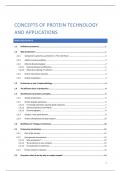
CONCEPTS OF PROTEIN TECHNOLOGY
AND APPLICATIONS
INHOUDSOPGAVE

1.1 Definition proteomics .......................................................................................................................... 1

1.2 Why proteomics? ................................................................................................................................ 1
1.2.1 Compared to genomics, proteomics is “the real thing” ....................................................................... 1
1.2.2 mRNA vs protein profiling ................................................................................................................... 2
1.2.3 More (6-8) proteins/gene .................................................................................................................... 2
1.2.3.1 Posttranslational modification ........................................................................................................ 2
1.2.3.2 Alternative splicing à isoforms ...................................................................................................... 3
1.2.4 Protein interaction networks ............................................................................................................... 3
1.2.5 Cellular localization ............................................................................................................................. 3

1.3 Proteomics as part of systemsbiology .................................................................................................. 4

1.4 The different faces of proteomics ........................................................................................................ 4

1.5 Identification of proteins: principles .................................................................................................... 5
1.5.1 Sample preparation ............................................................................................................................. 6

1.5.2 Protein peptide separation .................................................................................................................. 6
1.5.2.1 Increasing separation capacity (peak capacity) ............................................................................... 7
1.5.2.2 2D-Electrophoresis (2D-PAGE) ........................................................................................................ 7
1.5.2.3 Chromatography ............................................................................................................................. 8
1.5.3 Analysis: mass spectrometry ............................................................................................................... 9
1.5.4 Protein identification by data analysis .............................................................................................. 10

1.6 Workflows for ‘shotgun’ proteomics .................................................................................................. 10

2.1 Proteomics introduction .................................................................................................................... 11

2.1.1 Part of life sciences ............................................................................................................................ 11
2.1.2 from genome to proteome ................................................................................................................ 12
2.1.2.1 Why proteomics? .......................................................................................................................... 12
2.1.2.2 The proteome is very complex! ..................................................................................................... 12
2.1.2.3 From genome to proteome ........................................................................................................... 13
2.1.3 Protein variants in disease................................................................................................................. 13

2.2 Overview: what do we do with a complex sample? ........................................................................... 14


1

,2.3 Sample preparation steps .................................................................................................................. 15

2.3.1 Defining your research question ........................................................................................................ 16
2.3.2 Literature study ................................................................................................................................. 16

2.3.3 Sample collection............................................................................................................................... 17
2.3.3.1 Sample types ................................................................................................................................. 17
2.3.3.2 Sample degradation ...................................................................................................................... 17
2.3.4 Protein extraction – solubilization ..................................................................................................... 17
2.3.4.1 Protein extraction.......................................................................................................................... 17
2.3.4.2 Protein solubilization ..................................................................................................................... 20

2.3.5 Protein separation or purification ..................................................................................................... 22
2.3.5.1 Acetone precipitation .................................................................................................................... 22
2.3.5.2 Gelfiltration ................................................................................................................................... 22
2.3.5.3 Filtering.......................................................................................................................................... 22
2.3.5.4 SDS-PAGE....................................................................................................................................... 23
2.3.6 Reduction and alkylation ................................................................................................................... 23
2.3.7 Digestion............................................................................................................................................ 24
2.3.8 Peptides purification or selection ...................................................................................................... 25
2.3.9 Prefractionation ................................................................................................................................ 26

3.1 Introduction ...................................................................................................................................... 27

3.1.1 AA analyses and AA sequencing are two different techniques .......................................................... 27
3.1.2 What can Amino Acid Analysis (AAA) do for you? ............................................................................. 27

3.2 Sample Preparation for Amino Acid Analysis ..................................................................................... 28

3.3 AAA of proteins/peptides .................................................................................................................. 28

3.3.1 In general........................................................................................................................................... 28
3.3.2 Hydrolysis .......................................................................................................................................... 28

3.3.3 Separation of amino acids ................................................................................................................. 30
3.3.3.1 Paradigms ...................................................................................................................................... 30
3.3.3.2 Pre-column derivatization ............................................................................................................. 30
3.3.3.3 Post-column derivatization ........................................................................................................... 31
3.3.3.4 Pre- versus post-column derivatization ......................................................................................... 32

3.4 Amino acid sequencing ...................................................................................................................... 33
3.4.1 In general........................................................................................................................................... 33
3.4.2 Edman degradation ........................................................................................................................... 33
3.4.2.1 Insulin ............................................................................................................................................ 33
3.4.2.2 How does it work? ......................................................................................................................... 33
3.4.2.3 Cyclical estimation of yields .......................................................................................................... 34
3.4.2.4 Side products ................................................................................................................................. 34
3.4.2.5 Edman chemistry also disrupts AAs............................................................................................... 35
3.4.2.6 Positive aspects and drawbacks of Edman sequencing ................................................................. 35


2

, 3.4.3 Cleavage ............................................................................................................................................ 35
3.4.3.1 Chemical cleavage ......................................................................................................................... 35
3.4.3.2 Enzymatic cleavage ....................................................................................................................... 36

3.5 Carboxyterminal sequencing ............................................................................................................. 36
3.5.1 Carboxypeptidases ............................................................................................................................ 36
3.5.2 Chemical process ............................................................................................................................... 36

3.6 Single molecule sequencing ............................................................................................................... 37
3.6.1 Edman degradation ........................................................................................................................... 37
3.6.2 Microscale Edman degradation......................................................................................................... 37

4.1 Introduction ...................................................................................................................................... 39
4.1.1 Electrophoresis .................................................................................................................................. 39
4.1.2 2-dimensional gel electrophoresis ..................................................................................................... 39

4.2 Basic principles .................................................................................................................................. 39

4.2.1 In general........................................................................................................................................... 39
4.2.2 In practice .......................................................................................................................................... 40

4.3 First dimension: Isoelectric focusing (IEF)........................................................................................... 40
4.3.1 Isoelectric focusing ............................................................................................................................ 40
4.3.2 Protein charge ................................................................................................................................... 40
4.3.3 Creating a pH gradient for IEF ........................................................................................................... 41
4.3.3.1 Using carrier ampholytes .............................................................................................................. 42
4.3.3.2 IMMOBILIZED PH GRADIENTS ....................................................................................................... 42

4.3.4 2D-GE sample preparation ................................................................................................................ 43
4.3.5 Standard IP run for IEF and 2D-GE..................................................................................................... 44

4.4 Second Dimension: SDS-PAGE............................................................................................................ 44
4.4.1 SDS-PAGE ........................................................................................................................................... 44

4.4.2 Mass markers .................................................................................................................................... 45
4.4.3 PAGE matrix....................................................................................................................................... 46
4.4.3.1 Composition .................................................................................................................................. 46
4.4.3.2 Polymerization............................................................................................................................... 46
4.4.4 SDS-PAGE systems ............................................................................................................................. 46

4.5 Sample preparation ........................................................................................................................... 47
4.5.1 IEF preparation .................................................................................................................................. 47
4.5.2 Preparation of pH gradient gels ........................................................................................................ 47
4.5.3 IPG preparation for second dimension .............................................................................................. 47


3

, 4.6 Protein detection methods 2D-GE...................................................................................................... 48

4.6.1 Coomassie Brilliant Blue (CBB R-250) ................................................................................................ 48
4.6.2 Silver staining .................................................................................................................................... 49

4.6.3 SYPRO Ruby staining.......................................................................................................................... 49
4.6.4 2D-GE autoradiography..................................................................................................................... 49
4.6.5 Overview of staining options ............................................................................................................. 49

4.7 Analysis of 2D-gels ............................................................................................................................. 50
4.7.1 Image processing – digitalization ...................................................................................................... 50
4.7.2 Protein spot or ‘gradu’....................................................................................................................... 50

4.7.3 Complexities of 2D-GE ....................................................................................................................... 50

4.8 Differential in-gel electrophoresis (DIGE) ........................................................................................... 51
4.8.1 In general........................................................................................................................................... 51
4.8.2 Application ........................................................................................................................................ 51

4.8.3 Example ............................................................................................................................................. 52
4.8.4 DeCyder v. 6.5.................................................................................................................................... 52

4.9 2D-GE protein identification .............................................................................................................. 52

4.10 Take home points .............................................................................................................................. 52

5.1 Introduction ...................................................................................................................................... 53

5.2 Hydrophobic interactions .................................................................................................................. 53

5.3 Standard conditions for RP-HPLC ....................................................................................................... 54
5.3.1 Stationary phase................................................................................................................................ 54
5.3.2 Mobile phase ..................................................................................................................................... 55
5.3.2.1 Organic solvent .............................................................................................................................. 56
5.3.2.2 pH of mobile phase ....................................................................................................................... 58
5.3.2.3 Agents for ion-pairing .................................................................................................................... 58

5.4 Microcolumn Reverse Phase chromatography ................................................................................... 60
5.4.1 General .............................................................................................................................................. 60
5.4.2 Principle ............................................................................................................................................. 60

5.4.3 Adaptation of optimal flow rate ........................................................................................................ 61
5.4.4 Capacity and resolution of microcolumns ......................................................................................... 61
5.4.5 Adaptations of the HPLC system ....................................................................................................... 62

5.5 2D-LC ................................................................................................................................................. 63
5.5.1 Peak capacity and set-up of a 2D-LC ................................................................................................. 63


4

Voordelen van het kopen van samenvattingen bij Stuvia op een rij:

Verzekerd van kwaliteit door reviews

Verzekerd van kwaliteit door reviews

Stuvia-klanten hebben meer dan 700.000 samenvattingen beoordeeld. Zo weet je zeker dat je de beste documenten koopt!

Snel en makkelijk kopen

Snel en makkelijk kopen

Je betaalt supersnel en eenmalig met iDeal, creditcard of Stuvia-tegoed voor de samenvatting. Zonder lidmaatschap.

Focus op de essentie

Focus op de essentie

Samenvattingen worden geschreven voor en door anderen. Daarom zijn de samenvattingen altijd betrouwbaar en actueel. Zo kom je snel tot de kern!

Veelgestelde vragen

Wat krijg ik als ik dit document koop?

Je krijgt een PDF, die direct beschikbaar is na je aankoop. Het gekochte document is altijd, overal en oneindig toegankelijk via je profiel.

Tevredenheidsgarantie: hoe werkt dat?

Onze tevredenheidsgarantie zorgt ervoor dat je altijd een studiedocument vindt dat goed bij je past. Je vult een formulier in en onze klantenservice regelt de rest.

Van wie koop ik deze samenvatting?

Stuvia is een marktplaats, je koop dit document dus niet van ons, maar van verkoper BMWstudent19. Stuvia faciliteert de betaling aan de verkoper.

Zit ik meteen vast aan een abonnement?

Nee, je koopt alleen deze samenvatting voor €11,19. Je zit daarna nergens aan vast.

Is Stuvia te vertrouwen?

4,6 sterren op Google & Trustpilot (+1000 reviews)

Afgelopen 30 dagen zijn er 79223 samenvattingen verkocht

Opgericht in 2010, al 14 jaar dé plek om samenvattingen te kopen

Start met verkopen
€11,19  10x  verkocht
  • (0)
  Kopen