Neurological basis of the brain (GGZ2024)
Mapping the brain
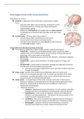
Cerebrum – largest part of the human brain, containing the cerebral
cortex.
o associated with higher order functioning, including the control
of voluntary behaviour, thinking, perceiving, planning and
understanding language
o divided into 2 hemispheres and bridged with the corpus callosum
hemispheres communicate with each other across the corpus
callosum.
Cerebral cortex – the outer layer of the cerebrum
o Consists of grey matter (containing neurons) and are
interconnected white matter (myelinated axons).
o The large furrows (groeven) are called fissures and the small ones
are called sulci (singular: sulces).
Cortical lobes and subcortical structures of the brain
The cerebrum is divided into 4 lobes: frontal, parietal, occipital and temporal:
o Frontal lobe - initiating and coordinating voluntary motor movements; higher
cognitive skills, such as problem solving, decision making, thinking, planning, and
organizing; and for many aspects of personality.
o Parietal lobe - integrating sensory processes from the body, , directing our attention,
and language.
o Occipital lobe - process visual information, including recognition of shapes and
colors.
o Temporal lobe - process auditory information, language and integrates information
from the other senses & complex visual processes.
Also memory through its hippocampus, and in (learned) emotional responses
through its amygdala.
Insular cortex – portion of the cerebral cortex folded deep within the lateral sulcus
o Involved in consciousness and plays a role in emotion and regulation of the body’s
homeostasis – empathy, perception, motor control, self/interceptiveawareness,
sensation of pain, and interpersonal experience.
Limbic system - arc shaped region of the cortex, located on both sides of the thalamus
o Involved in regulation of motivated behaviour (fleeing, feeding, fighting and sexual
behaviour), emotional life and formation and consolidation of memory (in
hippocampus and amygdala).
o Subcortical structures:
Thalamus - relaying most sensory information on to the cerebral cortex after
helping to prioritize it and regulation of consciousness, sleep and alertness
Hypothalamus - control center for appetites, defensive and reproductive
behaviors, and sleep-wakefulness – link between the nervous system to the
endocrine system, releasing hormones.
Cerebellum - helps control movement and cognitive processes that require
precise timing or attention & plays an important role in Pavlovian learning.
Basal ganglia - coordinate voluntary muscle movements, procedural
learning, routine behaviors or habits, reward and working memory.
1
, Directions and planes in the Nervous system
The vertebrate nervous system has 3 axes: anterior-posterior, dorsal-ventral, and medial-
lateral.
o Anterior: in front or towards to nose <->
posterior: to the back or toward to tail end
o Dorsal: toward the surface of the back or
to the top of the head (back)<-> ventral:
toward the surface of the chest or the
bottom of the head (front)
o Medial: close to the centre or toward the
midline of the body <-> lateral: to the side
or away from the midline.
Other useful terms:
o Proximal (nearest/ close) and distal (far)
o Ipsilateral: structures on the same side of the body; contralateral: structures on the
opposite sides of the body; unilateral: on one side of the body; and bilateral: both
sides of the body.
Planes (sections/ slices) to divide the brain: horizontal (superior and inferior), frontal (front
and back) and sagital (left and right).
The neuron
Neurons (cells within the nervous system) are the information processing and information
transmitting elements of the nervous system. Neurons specialized in the reception, conduction, and
transmission of information through electrical and chemical signals. A typical neuron consists of a cell
body/ soma, dendrites, an axon and terminal buttons.
Cell body/ soma: contains the nucleus, cytoplasm and many organelles. It give rise to
multiple dendrites (receiving signals) but only 1 axon (sending signals).
Axon / nerve fiber: extends from the cell body at a site called the axon hillock and gives rise
to many smaller branches before ending at the terminal buttons. Axons are covered with a
layered myelin sheath (fatty substance), this accelerates the
transmission of signals.
o An action potential starts at the axon hillock.
2


