GGZ2025 Neuropsychological Disorders vvanbeek
TASK 3 – DEVELOPMENT AND PLASTICITY
DEVELOPMENT OF CENTRAL NERVOUS SYSTEM
Source: Kolb & Whishaw (2015)
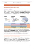
The developing brain originates as a three-part structure (equivalent to adult brain of fish or
reptiles). At later stages, the front and back components expand greatly and subdivide further,
with five regions in all. The three-part brain exists of the prosencephalon (front brain), which
is responsible for olfaction, the mesencephalon (middle brain) is responsible for vision and
hearing, and the rhombencephalon (hindbrain), which controls movement and balance. The
spinal cord is considered part of the hindbrain.
In mammals (part B of figure) the anterior prosencephalon forms the cerebral hemispheres,
known collectively as the telencephalon (endbrain). The posterior prosencephalon, known as
the diencephalon (between brain) includes the thalamus. Behind the mesencephalon, the
rhombencephalon develops further into the metencephalon (across brain) and the
myelencephalon (spinal brain, lower part of the brainstem). The human brain (part C of
figure) is more complex, and is mostly divided into forebrain, brainstem and spinal cord.
DEVELOPMENT OF THE HUMAN BRAIN
After fourteen days, the embryo consists of several sheets of cells with a raised area in the
middle; the primitive body. By three weeks after conception, it possesses a primitive brain (a
sheet of cells at one end). This sheet rolls up and forms the neural tube. By 7 weeks, the
embryo begins to resemble a miniature person, and about 100 days after conception, the brain
looks distinctly human. However, it does not begin to form gyri and sulci until 7 months.
1
, GGZ2025 Neuropsychological Disorders vvanbeek
In development, a series of changes take place in a fixed sequence (see table). This program
of development has two extraordinary features. First, nervous-system subcomponents form
from cells whose destination and function are largely predetermined. Second, development is
marked by an initial abundance of cells and connections, by apoptosis (genetically
programmed cell death).
Stages of brain development
1) Cell birth (neurogenesis, gliogenesis)
2) Cell migration
3) Cell differentiation
4) Cell maturation (dendrite and axon growth)
5) Synaptogenesis (formation of synapses)
6) Cell death and synaptic pruning
7) Myelogenesis (formation of myelin)
Deficits in genetic program, trauma, toxic agents or other factors may lead to errors in
development that contribute to deformities. The types of abnormal development are;
Types of abnormal development
Anencephaly Cerebral hemispheres, diencephalon and midbrain are absent.
Holoprosencephaly Cortex forms as a single undifferentiated hemisphere.
Lissencephaly Brain fails to form sulci and gyri.
Micropolygyria Gyri are more numerous, smaller, and more poorly developed.
Macrogyria Gyri are broader and less numerous than typical.
Microencephaly Development of brain is rudimentary, low intelligence.
Porencephaly Cortex has symmetrical cavities where cortex/white matter should be.
Heterotopia Displaced islands of gray matter, caused by aborted cell migration.
Callosal agenesis Entire corpus callosum or part is absent.
Cerebellar agenisis Parts cerebellum, basal ganglia or spinal cord are absent/malformed.
2
, GGZ2025 Neuropsychological Disorders vvanbeek
NEURON GENERATION
The neural tube has multipotential
neural stem cells, which have a great
capacity for self-renewal. In an adult,
these neural stem cells line the
ventricles, forming the
subventricular zone. Stem cells
have, besides lining the ventricles,
another function; they give rise to
progenitor (precursor) cells. The
progenitor cells also divide, but they
eventually produce nondividing cells
known as neuroblasts and glioblasts
that mature, respectively into
specialized neurons and glial cells.
Neural stem cells give rise to all of the many specialized brain and spinal cord cells.
Neurogenesis can continue into adulthood and even into senescence. This means that when
injury or disease causes neurons to die in an adult, perhaps the brain can de induced to replace
those neurons (which is unclear yet). The production of new neurons continuously suggests
that old neurons are dying.
CELL MIGRATION AND DIFFERENTIATION
The production of neuroblasts destined to form the cerebral cortex is largely complete by the
middle of gestation (4,5 months), whereas the cell migration to various regions continues for a
number of months, with some regions not completing migration until 8 months after birth.
During the last 4,5 months of gestation, the brain is most vulnerable to injury or trauma.
The brain can more easily cope with injury during neuron
generation than it can during cell migration and
differentiation. If neurogenesis is still progressing, the brain
may be able to replace its own injured cells or perhaps allocate
existing healthy cells differently. At the completion of general
neurogenesis, cell differentiation begins, in which neuroblasts
become specific types of neurons.
The cortex is organized into various areas that differ from
another cellular. How do cells know where these different parts
are located? The answer is that they travel along roads made of
radial glacial cells, each of which has a fiber extending from
the subventricular zone to the cortical surface (figure). The
cells from a given region of the subventricular zone only have
to follow the glacial road, and they end up in the right
location.
3


