Case 1 – The First Aid Groups
1) Which types of blood cells are there and what are their structure and function?
And how does this affect the coagulation?
First it would be important to note that there exist two types of immune systems, the innate immune system
(up to a few hours), which mainly plays a role in the coagulation, blood clotting and general healing of wounds
for this case, while it activates immediately and has been acquired via inheritance. And the adaptive immunity
which takes up to several days to start and makes specific antigens/cells to destroy the bacteria that entered
the body, and has to be acquired by the organism itself. The innate immune system kills of more general
bacteria and normally mainly protects the body due to physical barriers, if one of those barrier brakes the
innate immune system at first will try to fight of the foreign organisms entering the body.
The innate immune system consists out of epithelia cells, cells in the circulation and tissues and several plasma
proteins, these cells play complementary roles in preventing microbes to enter the body.
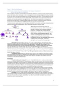
The Multipotential Hematopoietic stem cell
(Hemocytoblast) is the stem cell for all blood cells, it
separates its division into the common lymphoid
progenitor, of which its Natural Killer Cells and
Lymphocytes do not play a role in the innate immune
system, and the common myeloid progenitor. The
common myeloid progenitor is the precursor of the
megakaryocyte (and therefor thrombocytes),
erythrocytes, mast cells, and myeloblasts, which can be
subdivided later into basophils, neutrophils,
eosinophils and monocytes and macrophages.
- Erythrocytes are blood cells with a red colour, they do not contain a nucleus nor mitochondria and have a
biconcave disc (a donut shape with a closed hole); They do not contain most of the organelles so that they cannot use the
oxygen they transport themselves and have maximum carrying capacity for haemoglobin and thus oxygen. These
erythrocytes take up oxygen in the lungs and release carbon dioxide, travel via the blood vessels towards the remaining
parts of the body where they release this oxygen and take up carbon dioxide from these surrounding tissues.
The erythrocytes function for oxygen and carbon dioxide uptake and release is for 98% based on the haemoglobin found
within the erythrocytes; the haemoglobin takes up oxygen in a high oxygen environment causing the chemical equilibriums
to alter in such way that carbon dioxide will be released; in high carbon dioxide environments the opposite occurs (H2O +
CO2 ⇋ H2CO3 ⇋ H+ + HCO3- ⇋ H+ + HbO2 ⇋ HHb + O2). Haemoglobin molecules consist out of two α- and two β-chains
and four haemoglobin molecules.
- Thrombocytes/Platelets are parts of blood cells that are the first to form a plug at the site of injury in a blood
vessel, these platelets accumulate at the damaged site. This accumulation causes a temporary plug which prevents blood
from flowing out of the vessel, the platelets also secrete certain substances which signal more platelets to come and
Thromboxane A2 from their phospholipid bilayer.
Granulocytes
- The polymorphonuclear neutrophil (its name derived of the fact that its nucleus contains multiple
lobes) is the dominant white blood cell in the blood stream (50%-70%) and structure wise are closely related to
eosinophils and basophils; also granulocytes. The granules found within the neutrophil are mainly the primary
azurophil granules, which has the typical lysosomal morphology and filled with substances as myeloperoxidase
and nonoxidative antimicrobial effectors; and secondary specific granules containing lactoferrin and much of
the lysozyme. Neutrophils have a sort lifespan, from several hours up to several days.
These neutrophils are active in phagocytosing bacteria and are found in large amounts in the pus of wounds,
the neutrophils phagocytose multiple bacteria and are non-specific up to a certain point. Another reason that
pus has high levels of neutrophils is while they can capture multiple bacterial organisms, however their
lysosomal storage is limited, eventually causing the neutrophils to die while killing of the pathogens.
- Eosinophils (1%-4%), just like basophils and neutrophils originate from the myeloblast and mainly
roam through the blood itself, they contain granules filled with enzymes that can break down the cells walls of
pathogens, however these enzymes can also affect the cells of the host itself. Eosinophils have a bilobed
nucleus and can be stained with eosin.
1
, - Basophils (<1%), the third type of granulocyte, also have a high amount of granulocytes, filled with
fairly similar mediators as mast cells and are activated by IgE, in theory they could be seen as mast cells that
swim in the blood vessels, however their concentration in the human body is too low and actual defence
mechanisms are uncertain.
- Mature Mast cells will normally not be found in the blood stream, but only the tissue itself (mainly in
the skin, around the lips, blood vessels and nerves and other sites with potential injury), these mast cells are
filled with granules with acidic proteoglycans, which will be released if the mast cell’s high affinity IgE
membrane receptors are activated. Included in the acidic proteoglycans is histamine, which when released
causes surrounding blood vessels to vasodilate, and therefore increase the blood flow to an injured site and
allow more white blood cells to enter the tissue. Mast cells also release cytokines which signal leukocytes to
come to the site of injury itself.
Phagocytes
- Monocytes (12%), or Macrophages when they have left the blood stream, share a slightly similar
function with neutrophils, as in phagocytosing the bacteria encountered, but there the similarity ends.
Monocytes are the biggest white blood cells in the human body, and live much longer than neutrophils, on top
of that the macrophages are able to present pieces of the killed pathogen to T-lymphocytes to start creating
antigens. The macrophages of course have the same job, while they are essentially the same cell, however in
the tissue they quite often also phagocytose debris and dead cells of the human body itself.
- Both T and B Lymphocytes (20%-40%) are small cells who have a relative large nucleus, and only a
small amount of cytoplasm and poorly developed organelles. B Lymphocytes can be produced in bone marrow
tissue, originating the Common Lymphoid Progenitor, as where the T lymphocytes only can mature/be
produced in the Thymus. For both types of lymphocytes the same set of rules apply where they are actually
non-functional as mature lymphocytes in the blood stream, but can be activated by antigens in “Secondary
Lymphoid Organs” such as the spleen and the lymphoid nodes.
+ Both subtypes of lymphocytes can be further divided into general groupings of lymphocytes; T Lymphocytes
can majorly be subdivided into T-helper cells, which on their own do not kill pathogens, but by producing
cytokines or via direct contact they can activate other cells which do have a direct function in killing pathogens.
The second main group are the T-cytolytic cells which release granules filled with cytotoxic substances if in
contact with a foreign cell. Both types of T cells express T-Cell-Receptors, however the T-helper cells also
express CD4 next to that, which recognizes cells from the same organism via the MHC II molecules, as where T-
cytolytic cells recognize foreign cells by their MHC I Molecules/cells that need to be destroyed via the CD8
proteins on the cell membranes.
+ However not all foreign cells have a nucleus, and in general there are exceptions on the rules that nucleated
cells have MHC proteins, where the “Large Granular Lymphocytes” play a role, although better known as
Natural Killer Cells due to the fact that they are no lymphocytes actually, which kill organisms without MHCs.
+ B Lymphocytes on the other hand mainly find their function in killing pathogens by producing antibodies
which can bind onto these pathogens; the B cells can be subdivided into the multi-pathogen reactive antibody
producing B-1 cells and the mono-reactive antibodies of the B-2 cells.
(Naïve Lymphocytes have to be activated by encountering an antigen of a bacteria, without ever encountering
an antigen, either actively on its own or displayed by Antigen Presenting Cells, are rather functionless)-
Antigen-Presenting Cells (APCs) are cells that capture microbials and other antigen and display the
antigens to lymphocytes, which can use this information to produce antibodies, but the APCs also can provide
signals that stimulate differentiation and proliferation of lymphocytes. There are multiple Antigen-presenting
cells, such as;
+ Dendritic Cells play an important role in linking the innate immune system to the adaptive immune system,
which is based on the fact that they have phagocytotic capabilities and present parts/antigen of these captured
microbes to the T lymphocytes. They also have membrane receptors which can respond on microbe molecules
and start secreting cytokines themselves. These roles mainly are for the “Conventional Dendritic Cells”, the
Plasmacytoid DCs on the other hand can also produce soluble Type I Interferons and reorganize nucleic acids in
such ay that they can attack viruses on their own.
+ Follicular Dendritic Cells find and bind, and display, protein antigens for T and B lymphocytes to read, these
FDCs are no real DCs and mainly only found around the spleen and lymphoid nodes.
+ Also Macrophages and B Lymphocytes can present antigens to T lymphocytes.
2
, 2) How does the haemostasis work and its function? And
then mainly zoom into the coagulation process.
A damaged blood vessel needs to be repaired while still
allowing blood to flow through, however this blood flow
normally has to be decreased (by factors like
vasoconstrictors which lower the blood flow and
therefore pressure) whilst otherwise the platelet plaques
will wash away. Platelets do not contain a nucleus, the
cytoplasm however contains the normal mitochondria
and sooth endoplasmic reticulum, on top of that they
contain high amounts of granules filled with clotting
proteins and cytokines.
1) Vasoconstriction;
Damaged endothelial cells will release paracrine vasoconstrictors which narrow down the blood vessel, and
therefore decrease the blood flow through the vessel and the pressure within, allowing the platelet plug to be
formed/preventing it from flowing away.
2) Primary blockage by a platelet plug;
Under healthy/normal circumstances the endothelial lining
of a blood vessel prevents plaques from being formed by
forming a barrier between the blood and the collagen,
when a blood vessel ruptures however the endothelial cell
layer expose this collagen layer and platelets rapidly adhere
onto it.
Platelets adhere onto the integrins (membrane receptor
proteins linked to the cytoskeleton); these integrins also
activate the granular release of platelet factors, such as
serotonin, ADP and Platelet-Activating Factor (PAF).
Serotonin is a vasoconstrictor, as is thromboxane A2 which
can be made from the phospholipid bilayer of the platelets,
PAF along with ADP on the other hand increase the levels
of platelets by positive feedback.
3) Secondary haemostasis - Coagulation
The transformation from the platelet plug into the coagulation
plaque occurs via two pathways, which eventually intertwine with
each other, the so-called intrinsic and extrinsic pathway.
The Intrinsic Pathway begins when collagen or other activators are
exposed, which activates Factor XIIa, which then with the help of
Ca2+ ions activates Factor XIa, which activates Factor IXa which
then forms a complex with Factor VIII and activates Factor Xa.
The Extrinsic Pathway activates due to exposed Tissue Factors
(Factor III), which forms a complex with Factor XII and thereby also
activates Factor Xa.
Further down this cascade, in the Common Pathway, this pivotal
Xa works together with Va and PL and Ca2+ to catalyse the
prothrombin into active thrombin, and therefor creates the
‘thrombin burst’. This thrombin (Factor II) burst also signals
platelets, but its main function is the enhancement from the
deactivated Fibrinogen into the active Fibrin (Factor I). This fibrin
mashes together with other fibrin molecules, under the influence
of Factor XIII activated by thrombin itself and Ca2+ ions, which
cross-links all fibres and holds together the components of the
coagulation clot.
3
, These three steps however only form a
temporary fix, when the blood vessels slowly
starts repairing itself plasminogen activates
into plasmin, under the influence of thrombin
and tissue Plasminogen Activator (tPA), which
then slowly disintegrates the clot by
destabilizing fibrin (so-called fibrinolysis).
If it were not for chemical differences/barriers
between damaged vessels and undamaged
endothelial cells the blood vessels would clot
altogether; the endothelial cells themselves in
healthy circumstances prevent the platelets
from adhering to the underlaying collagen.
However also during the repair of damaged
parts of blood vessels the endothelial cells are
actively preventing the blood clot from
becoming oversized, anticoagulants disrupt
the intrinsic and extrinsic pathway in further
proximity of the damage by interfering with
certain factors; Heparin and Antithrombin III
for example block off activated IX, X, XI and
XII; Protein C inhibits V and VIII.
The blood clotting thus is prevented, in
healthy circumstances, as long as there is no
tissue damage to the endothelial lining of the
blood vessels. This prevention of blood
clotting highly decreases the chance on
thromboses, which can cause blood vessels to
block off or even more dangerously cause the
blood clot to shoot loose and enter the heart.
However blood clotting as read so far is
necessary to repair damaged blood vessels,
thus coagulation cannot be prevented in all
cases, but can be dissolved/ broken apart.
This prevention is not only caused while no
tissue factors can be bound, but also actively
by Antithrombin III and prostacyclin, which
block platelet aggregation and render
coagulation factors inactive.
When the blood vessel has been repaired, whilst the
blood clot itself is only a temporary fix and not a
permanent reparation, the endothelial cells (at least
I would guess so) start synthesizing and releasing the
nitric oxide and prostacyclin themselves again, and
Tissue Plasminogen Activators. This beautiful tPA
together with thrombin turns on plasminogen, and
thus creates the active plasmin; this active plasmin
breaks down the fibrin threads and cross links and
therefor destabilizes the blood clot, causing it to
dissolve; fibrinolysis. DAMPS
4


