Histology Lectures Chapter 4 (p.161 to p.172)
Epithelial and Gland Tissue
Cell biology (molecule – cell component – cell)
Histology (cell – tissue – organ)
Anatomy (organ – organ system)
Molecular biology/biochemistry (molecule – cell component)
Physiology (cell – tissue – organ – organ system)
Learning objectives for histology:
- Explain the relation between the shape and the function of cells
- Explain how cells together form a tissue, with the digestive system as an example
- Analyze histological preparations using light microscopy
- Draw the structure and organelles of the cell and explain their role in the cell function
There are around two hundred different cell types, which are organized in tissues. These tissues
can be classified in four basic types of tissue. The basic tissue types are functionally organized
in organs.
1. Epithelial Tissue
2. Connective Tissue
3. Muscle Tissue
4. Nervous Tissue
Cell types that are part of the same basic tissue type can look much different, but their
embryonic origin is the same (they share the same ancestor cell).
Germ layers:
Ectoderm: nervous tissue (skin), forms the exoskeleton.
Endoderm: digestive tissue (lung), forms the inner lining of organs.
Mesoderm: muscle tissue, connective tissue (derived from
mesenchyme cells), develops into organs.
* epithelia can be derived from either ectoderm, endoderm or
mesoderm layers.
Learning objectives for epithelial tissue:
- Able to recognize and name different types of epithelia
- Able to explain the relation between shape and function of different types of epithelia
- Able to name cellular characteristics and associated functions of epithelial cells
- Able to name intracellular connections and associated functions between epithelial cells
,Epithelial Tissue
An epithelium is a tissue existing of cells that are tightly connected to each other. It is derived
from one of the three primary germ layers (ectoderm, endoderm, mesoderm). There are two
types of epithelial tissue:
1. Covering epithelia (e.g. skin)
2. Glands (invagination of epithelial layers). During development, the epithelial layer will fold
and in that fold, cells will be able to secrete, for example, proteins
Classification of epithelia:
Embryonic origin: ectodermal, endodermal or mesodermal.
Morphology: shape of the cells.
Cell organization: number of cell layers (strata).
* epithelium is always on top of connective tissue.
Epithelial cell shapes – morphology
- Squamous epithelium (flat);
- Cuboidal epithelium (cubical, nuclei are centered)
- Transitional epithelium (changes between squamous and cuboidal epithelium, depends on
the stretching of the tissue, e.g. volume changes in the urine bladder)
- Columnar epithelium (elongated, elongated nuclei can have small structures on top)
Epithelium cell layers – cell organization
- Simple (one layer of cells)
- Stratified (multilayered)
Pseudostratified (a tissue that looks stratified, but is not actually stratified. The nuclei are at
different heights/levels, but all the cells have the same length)
Characteristics of epithelium
- No blood circulation
- Polarized: the epithelial cells have an apical (top) and basolateral side (bottom and side)
- Surface specializations (can be on either the apical or basolateral side)
- Presence of basal membrane (basal and reticular lamina, collagen matrix, proteoglycans)
Apical domain specializations
- Microvilli: increase surface area (plasma membrane), which can increase absorption of
components of the extracellular environment
* the cytoskeleton of microvilli is made of actin (microfilaments). A filament is an extension
of the cytoskeleton.
- Cilia: movement (of liquid on top of the cell), sensory function.
* five times larger than microvilli.
* the cilia have a cytoskeleton of microtubule (tubulin). The tubes can change their length
by contraction and relaxation. This makes the movement of cilia possible.
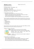
, Intercellular connection of epithelium cells
Tight junction
- Terminal web: the microvilli (actin
filaments) are connected to the cytoskeleton
via the terminal web; Adhesion
- Tight junction: cell – cell connection, in the belt
Terminal
upper part of the cell (above the terminal web
web), around the entire cell, zonula Button
occludens (prevents small molecules to go in desmosome
between the two cells), tight junction
proteins function like a zipper.
Function: prevents transport between cells,
membrane proteins of both cells are
compartmentalized (apical and basolateral)
to keep them separated, seals neighboring Hemidesmosome
cell together in an epithelial sheet to prevent (a)
Gap junctions
leakage of molecules between them;
- Adhesion belt: cell – cell connection, in the upper part of the cell, around the entire cell,
zonula adhaerens (adhesion between cells), made by transmembrane-linker proteins
(cadherins), the plasma membrane is, because of these adhesion molecules (CAMs: cell
adhesion molecules), a bit widened (intercellular), the adhesion molecules are connected
with actin to the terminal web (intracellular).
Function: joins an actin bundle in one cell to a similar bundle in a neighboring cell;
- Button desmosome: cell – cell connection, not around the entire cell, makes up the strongest
connection between cells; the dense area is connected to the cytoskeleton, it looks like the
adhesion belt, but the adhesion molecules (CAMs: cell adhesion molecules) are not made
up of actin; it is intracellularly connected with intermediate filaments (keratin, vimentin,
desmin, etc.).
Function: joins the intermediate filaments in one cell to those in a neighboring cell;
- Hemidesmosome: cell – tissue connection (binding to connective tissue underneath the
cell), half of the button desmosome, binding to fibers outside the cell (basal lamina: lamina
lucida and lamina densa), transmembrane-linker proteins (integrins) connect the cell
(intermediate filaments) with connective tissue (reticular fibers) underneath.
Function: anchors intermediate filaments in a cell to the basal lamina;
- Gap junction: cell – cell connection (nexus), gaps (pores in the membrane) by which the
cells can communicate with each other, transmembrane proteins that make up the gap
junction are called connexins (six connexins form one pore).
Function: intercellular transport (1.5nm), transport of ions, amino acids, molecules, certain
hormones, allows the passage of small water-soluble ions and molecules.
Basal domain specializations
- Hemidesmosomes;
- Basal lamina;
- Plasma membrane invaginations (basal labyrinth), increases the surface area, contains
mitochondria which provide energy needed for active transport in/out the cell.