Spierstelsel (BOOK)
50.5
Thin filaments are polymers of actin, coiled around one another. The same structure called
microfilaments function in cell mobility, one end elongates, while the other shortens.
Thick filaments are staggered arrays of myosin molecules.
Skeletal muscle
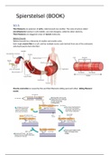
Skeletal muscle has a hierarchy of smaller and smaller units;
Each single muscle fiber is a cell, and has multiple nuclei, each derived from one of the embryonic
cells that fused to form the fiber.
Muscle contraction is caused by thin and thick filaments sliding past each other; sliding-filament
model.
, At rest, most muscle fibers contain only enough ATP for a few contractions, powering repetitive
contractions requires two other storage compounds; creatine phosphate and glycogen. The transfer
of a phosphate group from creatine phosphate to ADP can make up a new ATP molecule. This can
sustain contractions for about 15 seconds.
When glycogen is broken down to glucose, it releases lots of ATP, which is enough to sustain
contractions for nearly an hour aerobic! During activity, oxygen becomes limiting, and ATP is
instead generated by lactic acid fermentation, which generates much less ATP per glucose molecule,
and can sustain contraction for only 1 minute.
Muscle contraction is also caused by a power stroke from myosin, connected with actin. The only
struggle is that, in rest, tropomyosin (regulatory protein) and the troponin complex (set of additional
regulatory proteins) are bound to the actin strands of thin filaments. Tropomyosin covers the myosin
binding spot.
Motor neurons allow myosin and actin to bind, by triggering a release of Ca, which binds to the
troponin complex causing the myosin-binding sites on actin to be exposed. When Ca concentration
falls, the binding sites are covered and contraction stops.
Summary of contraction in a skeletal muscle;
1. The arrival of an action potential at the synaptic terminal of a motor neuron causes release
of the neurotransmitter acetylcholine. Binding of acetylcholine to receptor on the muscle
fibers leads to a depolarization that initiates an action potential. The action potential spreads
deep into the interior, following infoldings of the plasma membrane called transverse (T)
tubules. (which make close contact to the sarcoplasmic reticulum SR).
2. The action potential triggers changes in the SR, opening Ca-channels.
3. Ca ions stored in the SR flow through open channels into the cytosol (cytoplasm without
organelles, just the liquid).
4. Ca binds to the troponin complex.
5. Initiation of the muscle fiber contraction.
6. When motor neuron inputs stops, the filaments slide back relaxation. Relaxation starts
when proteins in the SR pumps Ca back into the SR from the cytosol.
7. When Ca concentrations decline, the regulatory proteins bound to the myosin binding sports,
blocking it.
Diseases
In Amyotrophic Lateral Sclerosis (ALS) motor neurons in the spinal cord and brainstem degenerate
and muscle fibers atrophy.
In myasthenia gravis, a person produces antibodies to the acetylcholine receptors of skeletal muscle.
Motor units
A motor unit consists of a single motor neuron and all the muscle fibers it controls. When a motor
neuron produces an action potential, all the muscle fibers in its motor unit contracts as a group. The
strength of the contraction depends on the amount of muscle fibers triggered.
Recruitment is the process in which more and more motor neurons are activated.
Some muscles (like the muscles that hold up the body and maintain posture) are continually
contracted. The nervous system alternates activation among the motor units, reducing the length of
time any set of fibers is contracted.
When the rate at which signals arrives is so high that the muscle fiber cannot relax at all between
stimuli, the twitches (single bout 100 ms) fuse into one smooth, sustained contraction tetanus.


