CLINICAL IMMUNOLOGY
Topic 1: Multiple Sclerosis
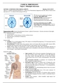
LECTURE 1: OVERVIEW OF MS CLINICAL ASPECTS Monday, 28/10/2019
Affects 2-3 million individuals globally 1:1000 in the Netherlands diagnosed with MS; affects women more
than men (2-3 : 1), disease of the young (20-40 yo)
CNS disease in the spinal cord; primary
pathology: INFLAMMATORY
DEMYELINATION of axonal myelin
disrupted axonal function due to
demyelinating lesions, detectable in MRI
Secondary pathological process: NON-
INFLAMMATORY DEGENERATION not so
visible in MRI; seen as atrophic lesions only if
it’s severe enough
Degeneration in MS: chronic demyelination of axons + gliosis of astrocytes + chronic activation of microglia
MRI localizations to look for MS lesions:
a. Periventricular
b. Juxtacortical – cortical (below or in the cerebral cortex)
c. Infratentorial (cerebellum & brainstem)
d. Spinal cord
Causes of MS:
a. Familial e.g. in monozygotic twins, 30% chance to develop MS among them, 2-5% increased risk if a
first-degree relative is affected
b. Environmental further away from equator, lower vit D status affect the risk of MS; viruses (EBV),
smoking (also associated with worse MS outcome), early obesity (e.g. since childhood)
c. Genes HLA region on chromosome 6 (also associated with many other autoimmune diseases, in
addition to MS); allelic variants in HLA-DRA locus is identified as risk factors for MS development –
more variants = more likely to develop MS
Symptoms of MS:
a. Optic neuritis (70% of all cases complains of having this; if one
has optic neuritis, there’s 50% chance to have MS diagnosis
later, thus require thorough MRI scan) decreased acuity,
central blurry-ness, loss of colour discrimination, retroorbital
pain
Eye movements disorder diplopia (d/t brainstem MS
lesion, usually in younger patients)
b. Lhermitte’s sign (if lesion in spinal cord) flexing the neck
(extend the spinal cord) causes the “electrical transmission”
down the back; MS suspicion if Lhermitte (+) in young patients,
Ddx for Lhermitte (+):
c. Motor symptoms (d/t demyelinating lesions down the motor
tract) loss of strength
d. Bladder-sexual dysfunction (one of the most common)
incontinence, sexual dysfunction
e. Psychological function disorders attention deficit, memory
loss, disturbed language functions, unable to multitask/slower
functioning
f. Fatigue (most common – albeit invisible): high impact, but
cause is unknown
, CLINICAL IMMUNOLOGY
Topic 1: Multiple Sclerosis
Symptoms arise due to inflammation within CNS semi-acute attack; origin: optic nerve, brain stem, spinal
cord. Mostly comes to the clinic with exacerbation of neurological symptoms keep on increasing over a few
weeks, then stagnates (complete/incomplete remission) before it comes up again
MS courses:
a. RRMS (relapsing remitting MS – most
common) inflammatory
predominance; unpredictable accidents
b. SPMS (secondary progressive MS)
degenerative predominance
c. PPMS (primary progressive MS)
typically older, male patients
How to diagnose MS:
Dx criteria: McDonald criteria (see below)
Monitored over 1 year
Diagnosis of MS has to be
reassured, both clinically &
radiologically, before
communicating to the patient
report this as CIS/clinically
isolated syndrome (symptoms +
w/o radiological evidence) or
RIS/radiological isolated
syndrome (radio + w/o
symptoms)
, CLINICAL IMMUNOLOGY
Topic 1: Multiple Sclerosis
Presence of oligoclonal bands in CSF if
oligoclonal bands concentration is high,
it is indicative for MS tx
Oligoclonal band: proteins that present
when there is inflammatory lesion of
CNS, when coupled with demyelinating
lesion evidence in MRI and/or MS
symptoms = MS
Other cases that allows presence of
oligoclonal band in CSF: … (?)
Has to make sure of both “dissemination in space & time”
a. Dissemination in space demyelinating lesions visible in 2 out of 4 localizations in MRI
b. Dissemination in time >1x relapse OR new lesions on follow-up MRI, contrast-enhancing lesion +
non-enhancing lesions at the same time
Contrast-enhancing lesion: active lesion/newly formed
Non-contrast-enhancing lesion: older lesion, but we don’t know how old
Tx aimed at RIS/CIS has to be weighed carefully
harm vs. benefit, although the risk of MS
development in those with RIS/CIS history is
very high
Diagnosis of PPMS has different Dx criteria:
a. Progression (+)
b. 2/3 the criteria above (time OR space OR
oligoclonal band)
MS follow up:
a. Clinical: frequency depending on
severity
b. Radiological (after 3 months)
c. Biochemical (biomarkers: neuro filament
light elevated when axon is broken
down, increased in serum & CSF)
Topic 1: Multiple Sclerosis
LECTURE 1: OVERVIEW OF MS CLINICAL ASPECTS Monday, 28/10/2019
Affects 2-3 million individuals globally 1:1000 in the Netherlands diagnosed with MS; affects women more
than men (2-3 : 1), disease of the young (20-40 yo)
CNS disease in the spinal cord; primary
pathology: INFLAMMATORY
DEMYELINATION of axonal myelin
disrupted axonal function due to
demyelinating lesions, detectable in MRI
Secondary pathological process: NON-
INFLAMMATORY DEGENERATION not so
visible in MRI; seen as atrophic lesions only if
it’s severe enough
Degeneration in MS: chronic demyelination of axons + gliosis of astrocytes + chronic activation of microglia
MRI localizations to look for MS lesions:
a. Periventricular
b. Juxtacortical – cortical (below or in the cerebral cortex)
c. Infratentorial (cerebellum & brainstem)
d. Spinal cord
Causes of MS:
a. Familial e.g. in monozygotic twins, 30% chance to develop MS among them, 2-5% increased risk if a
first-degree relative is affected
b. Environmental further away from equator, lower vit D status affect the risk of MS; viruses (EBV),
smoking (also associated with worse MS outcome), early obesity (e.g. since childhood)
c. Genes HLA region on chromosome 6 (also associated with many other autoimmune diseases, in
addition to MS); allelic variants in HLA-DRA locus is identified as risk factors for MS development –
more variants = more likely to develop MS
Symptoms of MS:
a. Optic neuritis (70% of all cases complains of having this; if one
has optic neuritis, there’s 50% chance to have MS diagnosis
later, thus require thorough MRI scan) decreased acuity,
central blurry-ness, loss of colour discrimination, retroorbital
pain
Eye movements disorder diplopia (d/t brainstem MS
lesion, usually in younger patients)
b. Lhermitte’s sign (if lesion in spinal cord) flexing the neck
(extend the spinal cord) causes the “electrical transmission”
down the back; MS suspicion if Lhermitte (+) in young patients,
Ddx for Lhermitte (+):
c. Motor symptoms (d/t demyelinating lesions down the motor
tract) loss of strength
d. Bladder-sexual dysfunction (one of the most common)
incontinence, sexual dysfunction
e. Psychological function disorders attention deficit, memory
loss, disturbed language functions, unable to multitask/slower
functioning
f. Fatigue (most common – albeit invisible): high impact, but
cause is unknown
, CLINICAL IMMUNOLOGY
Topic 1: Multiple Sclerosis
Symptoms arise due to inflammation within CNS semi-acute attack; origin: optic nerve, brain stem, spinal
cord. Mostly comes to the clinic with exacerbation of neurological symptoms keep on increasing over a few
weeks, then stagnates (complete/incomplete remission) before it comes up again
MS courses:
a. RRMS (relapsing remitting MS – most
common) inflammatory
predominance; unpredictable accidents
b. SPMS (secondary progressive MS)
degenerative predominance
c. PPMS (primary progressive MS)
typically older, male patients
How to diagnose MS:
Dx criteria: McDonald criteria (see below)
Monitored over 1 year
Diagnosis of MS has to be
reassured, both clinically &
radiologically, before
communicating to the patient
report this as CIS/clinically
isolated syndrome (symptoms +
w/o radiological evidence) or
RIS/radiological isolated
syndrome (radio + w/o
symptoms)
, CLINICAL IMMUNOLOGY
Topic 1: Multiple Sclerosis
Presence of oligoclonal bands in CSF if
oligoclonal bands concentration is high,
it is indicative for MS tx
Oligoclonal band: proteins that present
when there is inflammatory lesion of
CNS, when coupled with demyelinating
lesion evidence in MRI and/or MS
symptoms = MS
Other cases that allows presence of
oligoclonal band in CSF: … (?)
Has to make sure of both “dissemination in space & time”
a. Dissemination in space demyelinating lesions visible in 2 out of 4 localizations in MRI
b. Dissemination in time >1x relapse OR new lesions on follow-up MRI, contrast-enhancing lesion +
non-enhancing lesions at the same time
Contrast-enhancing lesion: active lesion/newly formed
Non-contrast-enhancing lesion: older lesion, but we don’t know how old
Tx aimed at RIS/CIS has to be weighed carefully
harm vs. benefit, although the risk of MS
development in those with RIS/CIS history is
very high
Diagnosis of PPMS has different Dx criteria:
a. Progression (+)
b. 2/3 the criteria above (time OR space OR
oligoclonal band)
MS follow up:
a. Clinical: frequency depending on
severity
b. Radiological (after 3 months)
c. Biochemical (biomarkers: neuro filament
light elevated when axon is broken
down, increased in serum & CSF)


