100% tevredenheidsgarantie Direct beschikbaar na je betaling Lees online óf als PDF Geen vaste maandelijkse kosten
logo-home
TEST BANK Dental Radiography: Principles and Techniques (6TH ED) by Joen Iannucci & Laura Jansen Howerton Chapters 1 - 35 | Complete €17,68
In winkelwagen

Tentamen (uitwerkingen)

TEST BANK Dental Radiography: Principles and Techniques (6TH ED) by Joen Iannucci & Laura Jansen Howerton Chapters 1 - 35 | Complete

 0 keer verkocht
  • Vak
  • Dental Radiography Principles and Techniques
  • Instelling
  • Dental Radiography Principles And Techniques
  • Boek

Dental Radiography: Principles and Techniques 6th Edition by Joen Iannucci & Laura Jansen Howerton Test Bank Stuvia Is Available For Download After Purchase. In Case You Encounter Any Difficulties with Download or want the document in a Different Format, Please Feel Free to Contact Me via Inbox. I ...

[Meer zien]
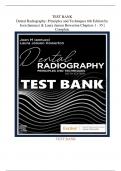
Voorbeeld 4 van de 372  pagina's

  • 7 november 2024
  • 372
  • 2024/2025
  • Tentamen (uitwerkingen)
  • Vragen en antwoorden
  • Dental Radiography Principles and Techniques
  • Dental Radiography Principles and Techniques
avatar-seller
TEST BANK
Dental Radiography: Principles and Techniques 6th Edition by
Joen Iannucci & Laura Jansen Howerton Chapters 1 - 35 |
Complete


TEST BANK

,Chapter 01: Radiation History
Iannucci: Dental Radiography, 6th Edition


MULTIPLE CHOICE

1. Radiation is defined as
a. a form of energy carried by waves or streams of particles.
b. a beam of energy that has the power to penetrate substances and record im
ageshadows on a receptor.
c. a high-
energy radiation produced by the collision of a beam of electrons with amet
al target in an x-ray tube.
d. a branch of medicine that deals with the use of x-rays.

ANSWER:C A
Radiation is a form of energy carried by waves or streams of particles. An x-
ray is a beam ofenergy that has the power to penetrate substances and record image shad
ows on a receptor.
X-radiation is a high-
energy radiation produced by the collision of a beam of electrons with ametal target in an
x-ray tube. Radiology is a branch of medicine that deals with the use of
x-rays.

DIF: Recall REF: Page 2 OBJ: 1
TOP: CDA, RHS, III.B.2. Describe the characteristics of x-radiation
MSC: NBDHE, 2.0 Obtaining and Interpreting Radiographs | NBDHE, 2.1 Principles of radiophysic
sand radiobiology

2. A radiograph is defined as
a. a beam of energy that has the power to penetrate substances and record im
ageshadows on a receptor.
b. a picture on film produced by the passage of x-rays through an object or body.
c. the art and science of making radiographs by the exposure of an image recepto
r tox-rays.
d. a form of energy carried by waves or a stream of particles.

ANSWER:C B
An x-
ray is a beam of energy that has the power to penetrate substances and record image shad
ows on a receptor. A radiograph is a picture on film produced by the passage of x-
rays through an object or body. Radiography is the art and science of making dental imag
es by theexposure of a receptor to x-
rays. Radiation is a form of energy carried by waves or streams of particles.

DIF: Comprehension REF: Page 2
OBJ: 1 TOP: CDA, RHS, III.B.2. Describe the characteristics of
x-radiation
MSC: NBDHE, 2.0 Obtaining and Interpreting Radiographs | NBDHE, 2.1 Principles of radiophysic
sand radiobiology

3. YourCpatient asked you why dental images are important. Which of the following is
thecorrect response?
a. An oral examination with dental images limits the practitioner to what is se
en clinically.
b. All dental diseases and conditions produce clinical signs and symptoms.

, c. Dental images are not a necessary component of comprehensive patient care.
d. Many dental diseases are typically discovered only through the use of den
talimages.
ANSWER:C D
An oral examination without dental images limits the practitioner to what is seen clinically.
Many dental diseases and conditions produce no clinical signs and symptoms. Dental images
are a necessary component of comprehensive patient care. Many dental diseases are typicall
y discovered only through the use of dental images.

DIF: Application REF: Page 2 OBJ: 2
TOP: CDA, RHS, III.B.2. Describe the characteristics of x-radiation
MSC: NBDHE, 2.0 Obtaining and Interpreting Radiographs | NBDHE, 2.5 General

4. The x-ray was discovered by
a. Heinrich Geissler
b. Wilhelm Roentgen
c. Johann Hittorf
d. William Crookes

ANSWER:C B
Heinrich Geissler built the first vacuum tube in 1838. Wilhelm Roentgen discovered the
x-
ray on November 8, 1895. Johann Hittorf observed in 1870 that discharges emitted from t
he negative electrode of a vacuum tube traveled in straight lines, produced heat, and result
ed in a greenish fluorescence. William Crookes discovered in the late 1870s that cathode r
ays were streams of charged particles.

DIF: Recall REF: Page 2 OBJ: 4
TOP: CDA, RHS, III.B.2. Describe the characteristics of x-radiation
MSC: NBDHE, 2.0 Obtaining and Interpreting Radiographs | NBDHE, 2.5 General

5. Who exposed the first dental radiograph in the United States using a live person?
a. Otto Walkoff
b. Wilhelm Roentgen
c. Edmund Kells
d. Weston Price

ANSWER:C C
Otto Walkoff was a German dentist who made the first dental radiograph. Wilhelm Roentg
en was a Bavarian physicist who discovered the x-
ray. Edmund Kells exposed the first dental radiograph in the United States using a live pers
on. Price introduced the bisecting technique in 1904.

DIF: Recall REF: Page 4 OBJ: 5
TOP: CDA, RHS, III.B.2. Describe the characteristics of x-radiation
MSC: NBDHE, 2.0 Obtaining and Interpreting Radiographs | NBDHE, 2.5 General

6. Current fast radiographic film requires
% less exposure time than the initial exposuretimes used in 1920.
a. 33
b. 98
c. 73

, d. 2

ANSWER:C D
Current fast radiographic film requires 98% less exposure time than the initial exposure ti
mesused in 1920.

DIF: Comprehension REF: Page 5
OBJ: 6 TOP: CDA, RHS, III.B.2. Describe the characteristics of
x-radiation
MSC: NBDHE, 2.0 Obtaining and Interpreting Radiographs | NBDHE, 2.5 General

7. Who modified the paralleling technique with the introduction of the long-cone technique?
a. C. Edmund Kells
b. Franklin W. McCormack
c. F. Gordon Fitzgerald
d. Howard Riley Raper

ANSWER:C C
C. Edmund Kells introduced the paralleling technique in 1896. Franklin W. McCormack r
eintroduced the paralleling technique in 1920. F. Gordon Fitzgerald modified the parallelin
gtechnique with the introduction of the long-
cone technique. This is the technique currently used. Howard Riley Raper modified the bi
secting technique and introduced the bite-wing technique in 1925.

DIF: Recall REF: Page 4 OBJ: 7
TOP: CDA, RHS, III.B.2. Describe the characteristics of x-radiation
MSC: NBDHE, 2.0 Obtaining and Interpreting Radiographs | NBDHE, 2.5 General

8. Which of the following is an advantage of digital imaging?
a. Increased patient radiation exposure
b. Increased patient comfort
c. Increased speed for viewing images
d. Increased chemical usage
ANSWER:C C
Patient exposure is reduced with digital imaging. Digital sensors are more sensitive to x-
rays than film. Digital sensors are rigid and bulky, causing decreased patient comfort. The i
mage from digital sensors is uploaded directly to the computer and monitor without the nee
d for chemical processing. This allows for immediate interpretation and evaluation. The ima
ge fromdigital sensors is uploaded directly to the computer and monitor without the need f
or chemicalprocessing.

DIF: Comprehension REF: Page 6
OBJ: 7 TOP: CDA, RHS, I.B.2. Demonstrate basic knowledge of di
gital radiography
MSC: NBDHE, 2.0 Obtaining and Interpreting Radiographs | NBDHE, 2.5 General

9. Which discovery was the precursor to the discovery of x-rays?
a. Beta particles
b. Alpha particles
c. Cathode rays
d. Radioactive materials

ANSWER:C C

Dit zijn jouw voordelen als je samenvattingen koopt bij Stuvia:

Bewezen kwaliteit door reviews

Bewezen kwaliteit door reviews

Studenten hebben al meer dan 850.000 samenvattingen beoordeeld. Zo weet jij zeker dat je de beste keuze maakt!

In een paar klikken geregeld

In een paar klikken geregeld

Geen gedoe — betaal gewoon eenmalig met iDeal, creditcard of je Stuvia-tegoed en je bent klaar. Geen abonnement nodig.

Direct to-the-point

Direct to-the-point

Studenten maken samenvattingen voor studenten. Dat betekent: actuele inhoud waar jij écht wat aan hebt. Geen overbodige details!

Veelgestelde vragen

Wat krijg ik als ik dit document koop?

Je krijgt een PDF, die direct beschikbaar is na je aankoop. Het gekochte document is altijd, overal en oneindig toegankelijk via je profiel.

Tevredenheidsgarantie: hoe werkt dat?

Onze tevredenheidsgarantie zorgt ervoor dat je altijd een studiedocument vindt dat goed bij je past. Je vult een formulier in en onze klantenservice regelt de rest.

Van wie koop ik deze samenvatting?

Stuvia is een marktplaats, je koop dit document dus niet van ons, maar van verkoper PureGold. Stuvia faciliteert de betaling aan de verkoper.

Zit ik meteen vast aan een abonnement?

Nee, je koopt alleen deze samenvatting voor €17,68. Je zit daarna nergens aan vast.

Is Stuvia te vertrouwen?

4,6 sterren op Google & Trustpilot (+1000 reviews)

Afgelopen 30 dagen zijn er 69052 samenvattingen verkocht

Opgericht in 2010, al 15 jaar dé plek om samenvattingen te kopen

Begin nu gratis
€17,68
  • (0)
In winkelwagen
Toegevoegd