Chapter 3 - An overview of nervous
system and muscles. Technical
investigations in neurology
Structure of the nervous system
Kuks, J. and Snoek, J., 2018. Textbook Of
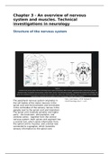
The peripheral nervous system originates in Clinical Neurology. pp.p.11 - p.25.
the cell bodies of the motor neurons in the
spinal cord and the brainstem and terminates
in the cell bodies of the sensory nerves in the
ganglia next to the spinal cord and brainstem.
The spinal cord (medulla spinalis) and higher
parts - the brainstem, diencephalon, and
cerebral cortex - together form the central
nervous system. Each spinal cord segment has
a ventral root, which sends information from
the spinal cord to muscles, and a dorsal root
connected to a ganglion, which transmits
sensory information to the spinal cord.
, The brain stem is divided from bottom (caudal) to top (cranial) into
The medulla oblongata (extension of spinal cord)
The pons (bridge crossed by various neural pathways)
The mesencephalon (midbrain)
The cerebellum, which plays a role in certain aspects of movement control, forms
an embryological unit with the pons. Further forward is the diencephalon, which
contains the thalamus (inner chamber), the hypothalamus (lower chamber), and the
hypophysis (undergrowth).
The hypothalamus is the coordination point for the autonomic (involuntary) nervous
system and the endocrine system.
The telencephalon (endbrain) comprises the cortex (cerebral cortex), the limbic
system and the basal ganglia. The cortex plays an important role in conscious
perception and action, the limbic system in episodic memory and emotion and the
basal ganglia in procedural activities (automatic and conscious and unconscious
motor function).
The CNS is surrounded by three membranes. The CSF enters the space between the
meninges around the brain and spinal cord, where it is discharged into the venous
system.
Two important main groups of tracts are the ascending tracts, which run up from
the spinal cord, and the descending tracts, which run down from the brain to the
spinal cord.
The muscle
Muscle spindles provide the CNS
with information on the state of
the muscle so that appropriate
action can be taken.
General description of membrane
potential - action potential
1. Action potential
travels along a motor nerve
to its endings on muscle
fibers
2. The nerve secretes a
small amount of acetylcholine
3. Acetylcholine opens
acetylcholine-gated channels through Kuks, J. and Snoek, J., 2018. Textbook Of
protein molecules floating in the Clinical Neurology. pp.p.11 - p.25.
membrane
4. Large quantities of sodium diffuse to the interior of the muscle fiber
membrane. This produces a local depolarization, which causes voltage-gated
channels to open, causing an action potential at the membrane.
5. The action potential travels along the muscle fiber membrane
6. The action potential depolarizes muscle membrane. The electricity of
the action potential in the sarcoplasm, the sarcoplasmic reticulum is activated
to release large quantities of calcium ions.
7. The calcium ions initiate a contraction between actin and myosin
filaments


