Chapter 15
Cell signaling
There are 3 major groups of cell singnaling:
1. Signaling through G-protein-coupled receptors
2. Signaling through enzyme-coupled receptors
3. Alternative signaling routes in gene regulation
Receptors are pharmaceutical targets.
Agonist: activates receptors
Antagonist: blocks or interferes with the receptor.
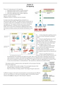
A simple intracellular signalling pathway activated by an
extracellular signal molecule. The signal molecule usually binds to
a receptor protein that is embedded in the plasma membrane of
the target cell. The receptor activates one or more intracellular
signalling pathways, involving a series of signalling proteins.
Finally, one or more of the intracellular signalling proteins alters
the activity of effector proteins and thereby the behaviour of the
cell. ----------------------------------------------------------------------->
(A) Contact-dependent signalling requires
cells to be in direct membrane-membrane
contact.
(B) Paracrine signalling depends on local
mediators that are released into the
extracellular space and act on neighbouring
cells. This are often the same kind of cells.
This also happens often in tissues.
(C) Synaptic signalling is performed by
neurons that transmit signals electrically
along their axons and release
neurotransmitters at synapses, which are
often located far away from the neuronal cell
body.
(D) Endocrine signalling depends on
endocrine cells, which secrete hormones into
the bloodstream for distribution throughout the body. Many of the same types of signalling molecules are
used in paracrine, synaptic, and endocrine signalling; the crucial differences lie in the speed and selectivity
with which the signals are delivered to their targets. The ligands of endocrine signalling are: hormones. There
is only a low concentration in the bloodstream. Their specificity is obtained via receptor binding.
The speed at which a response arises, also depends on the processes that take
place in the target cell.
- Fast: function of the protein changes. This can happen because of the
phosphorylation or because of a neurotransmitter.
- Slow: new proteins are being created. This changes the gene expression.
A different response in the same neurotransmitter can arise because two different
cells can react different on the same intracellular signal. An extracellular signal
molecule does not play a big part in the arising of a response.
,Extracellular signal molecules
the binding of extracellular signal molecules to either cell-surface or
intracellular receptors. Most signal molecules are hydrophilic and are
therefore unable to cross the target cell’s plasma membrane directly;
instead, they bind to cell-surface receptors, which in turn generate signals
inside the target cell.
Some small signal molecules, by contrast, diffuse across the plasma membrane
and bind to receptor proteins inside the target cell (either in the cytosol or in
the nucleus). Many of these small signal molecules are hydrophobic and poorly
soluble in aqueous solutions; they are therefore transported in the
bloodstream and other extracellular fluids bound to carrier proteins, from
which they dissociate before entering the target cell.
Each cell is programmed to respond to specific combinations of extracellular signals.
These signal molecules work in various combinations to regulate the behaviour of the cell. As
shown here, an individual cell often requires multiple signals to survive and additional signals
to grow and divide or differentiate. If deprived to appropriate survival signals, a cell will
undergo a form of cell suicide, known as apoptosis. The actual situation is even more
complex. Although not shown in this picture, some extracellular signal molecules act to
inhibit these and other cell behaviours, or even to induce apoptosis.
Different cells are specialized to respond to acetylcholine in different ways. Acetylcholine binds to similar receptor
proteins , but the intracellular signals produced are interpreted differently in cells specialized for different functions.
Cell-surface receptors relay signals via intracellular signalling molecules.
(A) A protein kinases covalently adds a phosphate from
ATP to the signalling protein, and a protein phosphatase
removes the phosphate. Although not shown in this
picture, many signalling proteins are activated by
dephosphorylation rather than by phosphorylation.
(B) A GTP-binding protein is induced to exchange its
bound GDP for GTP, which activates the protein; the
protein then inactivates itself by hydrolysing its bound GTP
to GDP.
The regulation of a monomeric GTPase. GTPase-activating proteins
(GAP’s) inactivate the protein by stimulating it to hydrolyse its bound
GTP to GDP, which remains tightly bound to the inactivated GTPase.
Guanine nucleotide exchange factors (GEF’s) activate the inactive
protein by stimulating it to release its GDP; because the concentration
of GTP in the cytosol is 10 times greater than the concentration of GDP,
the protein rapidly binds GTP and is thereby activated.
Hydrolysing GTP: GTPase.
The RAS protein is an exception of GTP binding.
, (A) In this simple signalling system, a
transcription regulator is kept in an
inactive state by a bound inhibitor
protein. in response to some upstream
signal, a protein kinase is activated and
phosphorylates the inhibitor, causing its
dissociation from the transcription
regulator and thereby activating gene
expression.
(B) This signalling pathway can be
diagrammed as a sequence of four steps,
including two sequential inhibitory steps
that are equivalent to a single activating
step.
Intracellular signalling complexes form at activated receptors.
(A) A receptor and some of the intracellular
signalling proteins it activates in
sequence are preassembled receptor by
a large scaffold complex.
(B) A signalling complex assembles
transiently on a receptor only after the
binding of an extracellular signal
molecule has activated the receptor;
here, the activated receptor
phosphorylates itself at multiple sites,
which than act as docking sites for
intracellular signalling molecules.
(C) Activation of receptor leads to the increased phosphorylation of specific phospholipids (phosphoinositides) in
the adjacent plasma membrane; these then serve as docking sites for specific intracellular signalling proteins,
which can now interact with each other.
Slow and rapid responses to an extracellular signal. Certain types of signal-
induced cellular responses, such as increased cell growth and division, involve
changes in gene expression and the synthesis of new proteins; they therefore
occur slowly, often starting an hour or more after the signal is received. Other
responses (such as changes in cell movement, secretion, or metabolism) need
not involve changes in gene transcription and therefore occur much more
quickly, often starting in seconds or minutes; they may involve the rapid
phosphorylation of effector proteins in the cytoplasm, for example. Synaptic
responses mediated by changes in membrane potential are even quicker and can
occur in milliseconds. Some signalling systems generate both rapid and slow responses as shown here, allowing
the cell to respond quickly to a signal while simultaneously initiating a more long-term, persistent response.


