100% tevredenheidsgarantie Direct beschikbaar na betaling Zowel online als in PDF Je zit nergens aan vast
logo-home
Summary all lectures nutrition in health and disease (master healthsciences) €3,89
In winkelwagen

Samenvatting

Summary all lectures nutrition in health and disease (master healthsciences)

 84 keer bekeken  10 keer verkocht

dit document bevat alle 10 de hoorcolleges van het vak nutrition in health and disease. Zowel de powerpoint als gesproken tekst van de docent is uitgewerkt, waardoor het document je een volledig overzicht geeft van de leerstof. this document contains all 10 lectures of the nutrition in health and...

[Meer zien]
Voorbeeld 7 van de 59  pagina's

  • 7 oktober 2020
  • 59
  • 2020/2021
  • Samenvatting
Alle documenten voor dit vak (1)
avatar-seller
an1997
Lectures nutrition in health and disease NHD

Lecture 2: (patho)physiology of GI tract (tue 8 sep 2020)
In this lecture, Sophie Rietveld, MSc (dietician Department of Surgery, Amsterdam UMC) will explain the
(patho)physiology of the gastrointestinal tract at advanced level. Special focus will be on the role of nutrition
and the dietetic treatment

The gastrointestinal tract in relation to Nutrition and dietetics

Topics:
- Physiology of the gastrointestinal tract in health and disease
- Biochemical processes and pathways involved in the digestive metabolism of the principle macro nutrients
and micronutrients
- Causes and (nutritional) consequences of diseases (e.g. cancer) of the GI tract
- Role of nutritional care around surgery

Health and disease:
Metabolism is a term that is used to describe all chemical reactions involved in
maintaining the living state of the cells in an organism. In case of illnesses the
processes are disrupted, for example in catabolism where the cells are broken
down, there is now balance anymore.

GI-tract: in this lecture we will focus on the esophagus, stomach
and pancreas
• Mouth
• Esophagus
• Stomach
• Pancreas
• Bile tract
• Small intestine
• Colon

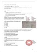
Important to know is that there is a difference between digestion and absorption. Digestion is the breakdown
of nutrients and absorption is the uptake of nutrients. In the table you can see the different organs and their
function in digestion and absorption. For example, the stomach has an important role in the digestion of
nutrients but has no role in the absorption of nutrition.

Mouth: The digestion of nutrition starts in the mouth.
• Chewing and mixing food with saliva
• Secretion of saliva -> contains enzymes (amylase)
• Amylase starts breakdown of carbohydrates
Production of saliva per day 1-1,5 L

Esophagus:
• Function: transport of food from mouth to stomach
• Height: 30-40 cm
• Bridging between pharynx (throat) and stomach
During swallowing, the epiglottis closes of the respiratory system and prevents
choking

• Structure: muscle- connective tissue – mucous
• Circular muscle – longitudinal muscle
• Connective tissue layer
• In connective tissue layer: mucus forming glands


1

,The esophagus consists out of 3 layers: muscular layer, connective tissue layer and mucous layer. The muscular
layer creates a wave like muscular contraction, this is called peristalses, as a result food is pushed down the
esophagus. Food is moving downwards by gravity.

Diseases of the esophagus:
• Esophagitis: inflammation of the esophagus
• Barrett’s disease: a, part of the esophagus is lined with different tissue than normal. A Barrett's esophagus
is caused by the return of heartburn into the esophagus for years. This is called reflux.
• Achalasia: problems with peristalsis; the muscle movement in the lower esophagus is reduced. As a result,
the sphincter between the esophagus and the stomach cannot relax properly. The sphincter can no longer
open properly due to this cramping. Food cannot pass through the esophagus properly and accumulates
above the sphincter.
• Carcinoma: cancer. The 8th most common cancer in NL. Risk factors: reflux and obesity, smoking, alcohol
and nutrition. Patients often report dysphagia (difficulty in swallowing)

Gastrointestinal tract after esophagectomy:
After radiation and chemotherapy there is surgery were a part of the
stomach and esophagus will be removed and the stomach is pulled up
like a tube. Esophagectomy followed by gastric tube reconstruction is
the surgical treatment of choice for patients with esophageal cancer
Tube stomach: esophagus removed + transition from esophagus to
stomach, remnant = stomach stretched as new esophagus. With a tube stomach you have no sphincter
function.

Complaints after esophagectomy: Due to the removal of the esophagus and the creation a gastric tube the
storage function of the stomach is reduced.
• Malnutrition and weight loss
• suboptimal intake of multiple vitamins and minerals
• Decrease in quality of life
• Fatigue
• Eating problems

Top 5 eating complaints after esophagectomy:
Patients who underwent a gastric tube reconstruction experience nutrition
related symptom during the first year after surgery. After 3 months, patients
get used to the new autonomy and complaints. when the stomach empties to
quickly, dumping syndrome may occur. The emptying of the stomach is
controlled by the pyloric sphincter. When the sphincter does not work
properly, food can enter the small intestine too quickly or in large portions.

◦ Early dumping: large amount of undigested food attracts water
can occur after half an hour after a meal. These symptoms arise because food attracts fluid in the
intestine. This fluid does not come from the intestine but is attracted from the bloodstream and is mixed
with food in the small intestine. This created a full feeling, cramps and diarrhea. Because less flood
circulates in the blood stream, a drop in blood pressure results, the hart pumps the blood around with less
pressure, making the pulse faster. By doing this, dizziness and weakness but also cramps and a full feeling
may occur
◦ Late dumping: sugars are absorbed into the blood too quickly
occurs later within one and a half or two hours after a meal. As a result of a reduced storage function of
the stomach, the digestion of the food is faster and the pancreas produces more insulin in a shorter time,
it is a hormone that ensures that glucose can absorbed from the bloodstream into the body cells. This
insulin production results in a drop of blood sugar, which creates diabetes like complaints such as shaking,
sweating, weakness. Over time, due to adaptation of the body, these symptoms become less severe
automatically but can hold from several hours.
◦ Some people suffer from both, early and late dumping but they can also occur separately.


2

,Stomach:
• Temporary storage food (primary function)
• Gastric fluid contains pepsin -> protein digestion
• Hydrochloric acid production
• Grinding and mixing food with gastric juice
• Gradually release of food to small intestine

The upper part (fundus and corpus) of the stomach stores food. Little by little the
stomach travels the food to its lower part. The food goes to the pyloric sphincter which allows small amounts
of chyme to pass to the small intestine and then close again. The stomach produces 1-2 L gastric fluid.

pH:
the hydrochloric acid is aggressive because of the acidity. Because of this it prevents bacterial growth
and kills bacteria that comes with the food. To protect the stomach itself against gastric fluid and
enzymes, the cells of the walls produce mucous. The saliva enzymes do not work in the acidity of the
stomach.

Diseases of the stomach:
• Gastritis: inflammation of the gastric mucosa.
• Stomach ulcer: occur when the thick layer of mucus that protects your stomach from digestive
juices is reduced. This allows the digestive acids to eat away at the tissues that line the stomach,
causing an ulcer
• Carcinoma: cancer

Role of the stomach in the absorption of vitamin B12:
• Vitamin B12 is present in meat, liver, eggs, cheese and milk
• Absorption of B12 in the ileum
• Resorption only possible after connection with intrinsic factor
• Intrinsic factor production in parietal cells fundus/corpus stomach

Potential causes of vitamin B12 deficiency:
• Insufficient bound of consumed vitamin B12 to intrinsic factor
• Suboptimal vitamin B12 absorption in the small intestine
• Insufficient vitamin B12 food consumption

• Autoimmune gastritis= reduced production of gastric juice: HCL, intrinsic factor, and pepsinogen
• Atrophic gastritis: process of chronic inflammation of the gastric mucosa of the stomach
• Lack intrinsic factor in stomach
• Total gastrectomy

Communication in the GI tract:
How does the body know what is necessary for digestion?

Hormone regulation of GI tract- stomach:
- Coordinates the digestive and absorptive processes. The hormone gastrin regulates
the acidity (pH) in the stomach, secreted by the cells of the stomach wall. The entry
of food into the stomach stimulates the cells to release gastrin, which stimulates
HCL. When the pH 1,5 is reached. The acid itself turns of the gastric production and
as a result the HCL production will stop.
- Gastrin, released from the stomach wall in response to food in the stomach;
stimulates the stomach to release HCL which ensures the pH of the stomach.


3

,Biliary tract:
- Refers to the liver, gallbladder and bile duct and how they
work together to create, store and secrete bile.
- The bile duct and the pancreatic duct come together in the
duodenum= papilla of Vater

Liver:
1. Metabolic fuction:
a. Role in metabolism of carbohydrates, fats, proteins
-> foodconcersion
b. Detoxification: purifying the blood from waste from the metabolism and harmful/toxic substances
c. Hormone breakdown
d. Storage and distribution
2. Secretoy function:
a. Production of bile
3. Vasculair function

The most important function of the liver related to food are the metabolic and secretory function. The liver
plays an important role in food conversion and storage, for example the storage of glucose in glycogen

Production bile juice:
The liver produces bile which is concentrated and stored in the gallbladder which secretes the
bile in the duodenum. Bile will be released when eating fatty foods. Per day, the body makes
400-500 ml bile.

Bile juice:
• Bile is an emulsifier that brings fat into a suspension in water so that enzymes can break
them down(Bile is not an enzyme) (this function can be compared to micelles and soap)
• After digestion reabsorbed by intestinal cells
• Return to liver via hepatic portal vein, bile bound to fiber will be excreted via feces.
• Brown color of stool due to bile

Pancreas:
• Structure: head, body, tail
• Position: behind stomach and part duodenum
• Length: 12-15 cm
• Weight: 70-100 g

Endocrine and exocrine function of the pancreas:
• Endocrine function: (mostly in the tail) -> hormones
o Insulin and glucagon
• Exocrine: (mostly in the head) -> enzymes
o +- 1 to 1,5 L pancreatic juice per day
o Enzymes: amylase (carbohydrate), lipase (fat) and proteases (protein)
o Bicarbonate to neutralize stomach acid


4

,Communication: hormonal secretion:
◦ Secretin, released from the duodenal wall in response to the
presence of "acidic" chyme in the duodenum; When secretin
reaches the pancreas, it stimulates the pancreas to release juice
with bicarbonate. In addition, secretin inhibits acid secretion in
the stomach. This is done by inhibiting the cells that produce
gastrin.
◦ Cholecystokinin CCK, is released by the wall of the small intestine
in response to the presence of fat (and protein) in the small
intestine
◦ Trypsinogen is converted by enterokinase into the active trypsin

(Also, see nutrition and research lecture 2)

Causes pancreatic cancer:
• Multi factorial
• Hereditary
• Is more common in people who
o >65 year
o Suffer from chronic pancreatic inflammation
o Smoke
o Have excessive alcohol consumption
• Increasing risk by low fiber diet
• Reducing risk by large vegetable consumption

The position of the tumor is often located in the head (75%), body (15%), tail (10%).

Symptoms in pancreatic cancer:
• Phase 1: weight loss, abnormal bowel habits, loss of appetite
• Phase 2: pain (stomach/back), jaundice, discolored feces/dark urine
• Phase 3: itching, nausea, vomiting, intestinal bleeding, closure, swelling belly

Complaints exocrine pancreas dysfunction:
• Malabsorption: when the tumor is located in the head, in can be an obstruction for pancreatic juice and
bile because it cannot be digested.
• Abnormal defecation: lack of bile results in white color.
o Frequency
o Consistency
o Color
• Body weight loss: loss of energy because of problems with fat digestion/absorption

Whipple surgery:
The most common surgery for pancreatic cancer. The head of the pancreas next to the gallbladder and a part
of the duodenum is removed. Many patients suffer from exocrine pancreas disfunction.

Pancreatic enzyme supplementation:
• In particular, pancreatic enzymes
• Requirement depends on (fat) intake
• Main and in-between meals
Purpose: prevention of malnutrition and reduction of complaints

How to measure:
• Visually check
• Fecal elastase:
• Average food- and fat intake and average fat in stool of 3-4 days


5

,It is possible that the remaining part of the pancreas produces pancreatic enzymes, despite the head being
removed, the most important part for making pancreas enzymes. In practice a standard dose is given. If
necessary, food intake is changed. A more objective measurement to measure exocrine function is to
determine the fecal elastase; a protein splitting enzyme that contributes to the breakdown and digestion of
protein. It is exclusively produced in the pancreas. In contrast to other protein splitting enzymes from the
pancreas, elastase remains intact after passing through the intestine. However, fecal elastase can be influenced
by intestinal transit time. Rapid intestinal transit can cause false-positively low fecal elastase. The most reliable
measurement for (fat) malabsorption is the average food and fat intake and measuring the average fat in stool
of 3-4 days.

Background BOMB-calorimetry:
• 4 days (weighted) diary of food intake
• (last) 3 days feces collection
• Burn reaction (measuring heat= energy)

By measuring nutrients in feces and by calculating the intake of nutrients, the nutritional uptake cam be
determined and we can determine whether patients have malabsorption. This measurement is called BOMB,
feces is burnt to measure energy.

Small intestine: duodenum-jejunum-ileum
• small intestinal fluids (1,5-2L per day) provide a last step in digestion/breakdown of carbohydrates and
proteins
• increase of absorption surface by villi and microvilli -> absorption.
• The majority of the nutrients are absorbed in the small intestine.

Absorption:
Trace elements are mainly absorbed in the duodenum. Absorption of
carbohydrate, fats and proteins and vitamin C and B takes place in the
jejunum. Vitamin B12 can only be absorbed at the end of the ileum.
In the colon: water, electrolytes (e.g. sodium and potassium) and vitamin
K are absorbed.

Colon:
• Large instestine
• Approximately 1,5 m
o Coecum
o Colon ascendens
o Colon transversum
o Colon descendens
o Rectum

The role of nutrition in surgery

Surgery and nutrition:
• Surgery -> stress and inflammatory response
• Nutrition: restore protein mass and/or an adequate stress response

Surgery leads to inflammation and metabolic stress response. To achieve healing and recovery, a metabolic
response is necessary. However, this metabolic response causes catabolism, leading to a loss of muscle mass.
Therefore, it is essential that patients have adequate nutritional therapy. Especially when the metabolic stress
response is present over a longer period of time.
The loss of muscle is a short- and long-term burden for recovery. Nutritional therapy and physical exercise aim
to restore or maintain muscle mass. However, in severally malnourished patients, nutritional therapy is not
sufficient enough to maintain or restore muscle mass but it provides an adequate metabolic stress response to
increase the changes of recovery.


6

, ERAS: Better and faster recovery, even after major operations. The program consists out of multiple
components before and after surgery to minimize stress and return function.

Early oral feeding is the preferred mode of nutrition for surgical patients. Avoidance of any nutritional therapy
bears the risk of underfeeding during the postoperative course after major surgery. Considering that
malnutrition and underfeeding are risk factors for postoperative complications, early enteral feeding is
especially relevant for any surgical patient at nutritional risk, especially for those undergoing upper
gastrointestinal surgery. The focus of this guideline is to cover nutritional aspects of the Enhanced Recovery
After Surgery (ERAS) concept and the special nutritional needs of patients undergoing major surgery, e.g. for
cancer, and of those developing severe complications despite best perioperative care. From a metabolic and
nutritional point of view, the key aspects of perioperative care include:
◦ integration of nutrition into the overall management of the patient
◦ avoidance of long periods of preoperative fasting
◦ re-establishment of oral feeding as early as possible after surgery
◦ start of nutritional therapy early, as soon as a nutritional risk becomes apparent
◦ metabolic control e.g. of blood glucose
◦ reduction of factors which exacerbate stress-related catabolism or impair gastrointestinal function
◦ minimized time on paralytic agents for ventilator management in the postoperative period
◦ early mobilization to facilitate protein synthesis and muscle function

See slide 53-60.

Example patient:
Cancer, weight loss, tube feeding. after surgery he gained 4 kg and has oral intake again. Still at risk of
malnutrition? Does he still need nutritional support? Yes, surgery leads to inflammation and metabolic stress
response which will break down the muscle proteins. Muscle mass is essential for recovery.

Diagnosis of malnutrition (GLIM criteria):
- measurable (phenotypic) criteria: weight loss, low BMI and low muscle mass
- causal (etiological) criteria: a reduced intake, increased losses or acute or chronic disease / inflammation
According to this definition, we speak of malnutrition when someone meets at least 1 of the 3 phenotypic
criteria in combination with at least 1 of the 3 etiological criteria.6


7

Voordelen van het kopen van samenvattingen bij Stuvia op een rij:

Verzekerd van kwaliteit door reviews

Verzekerd van kwaliteit door reviews

Stuvia-klanten hebben meer dan 700.000 samenvattingen beoordeeld. Zo weet je zeker dat je de beste documenten koopt!

Snel en makkelijk kopen

Snel en makkelijk kopen

Je betaalt supersnel en eenmalig met iDeal, creditcard of Stuvia-tegoed voor de samenvatting. Zonder lidmaatschap.

Focus op de essentie

Focus op de essentie

Samenvattingen worden geschreven voor en door anderen. Daarom zijn de samenvattingen altijd betrouwbaar en actueel. Zo kom je snel tot de kern!

Veelgestelde vragen

Wat krijg ik als ik dit document koop?

Je krijgt een PDF, die direct beschikbaar is na je aankoop. Het gekochte document is altijd, overal en oneindig toegankelijk via je profiel.

Tevredenheidsgarantie: hoe werkt dat?

Onze tevredenheidsgarantie zorgt ervoor dat je altijd een studiedocument vindt dat goed bij je past. Je vult een formulier in en onze klantenservice regelt de rest.

Van wie koop ik deze samenvatting?

Stuvia is een marktplaats, je koop dit document dus niet van ons, maar van verkoper an1997. Stuvia faciliteert de betaling aan de verkoper.

Zit ik meteen vast aan een abonnement?

Nee, je koopt alleen deze samenvatting voor €3,89. Je zit daarna nergens aan vast.

Is Stuvia te vertrouwen?

4,6 sterren op Google & Trustpilot (+1000 reviews)

Afgelopen 30 dagen zijn er 59804 samenvattingen verkocht

Opgericht in 2010, al 15 jaar dé plek om samenvattingen te kopen

Start met verkopen
€3,89  10x  verkocht
  • (0)
In winkelwagen
Toegevoegd