HC 8 Sensorimotor function and anomalies
Chapter 11
This is what we will talk about today.
(reads what’s on the slide)
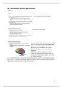
Here you have the brain with the cerebrum so the
fore lobe, with the four lobes: the temporal lobe,
occipital lobe, parietal lobe, frontal lobe. And
below there is the cerebellum, that is also a
structure that plays an important role in our
movement.
Other regions that are involved in our motor
system are subcortical basal ganglia, within the
cerebrum. And that helps to produce movement,
for example grasping.
And the cerebellum helps to regulate the timing and accuracy of movement. It calculates something
like an error, so If we want to grasp something and we miss is, then the cerebellum comes involved in
regulating this movement and correcting the movement.
1
, And you have already
seen this figure in the first
lecture. So we have
different sensory
pathways. We have
afferent somatosensory
pathways where
information travels from
the body inward via the
somatic nervous system.
And we have movement
information that travels
out of the CNS via a
parallel. And that is the efferent motor pathway. And you see here this color coding. So we have
sensory endings, for example if you step on a pin then the information the pain sensation travels
inwards and these are the efferent pathways. And we have our information that travels from the
brain outwards, and these motor pathways are called afferent.
This is shown in more
detail here in this
figure. How Is it
organized. If we start
with one, we have our
visual information.
And this is required to
locate a target so if
you want to grasp a
cup of coffee here,
then you first need to
visually locate the
target. And in your
frontal lobe, that is the
second part here, our
motor area plans our
movements. So the
reaching component is
planned in the frontal-lobe. And then we have the spinal cord, that is number 3, the spinal cord
caries the information to the hand. And then we have our motor neurons that carry the message to
the muscles of the hand and the forearm. And then important, number 5, in our sensory receptors
that we have in our fingers, they make contact with a cup of coffee and send the message to the
sensory cortex, saying that the cup has been grasped. Or feeling pain sensation if the cup is too hot.
So this information is then carried backwards via the sensory nerve, and the spinal cord carries the
sensory information to the brain, either that the cup has been successfully grasped or that the cup is
too hot or that the cup has been missed. And then in the middle structure of the brain we have our
basal ganglia that judges the grasp. So judges whether the grasp has been successful, whether the
force of the fingers is strong enough to really hold the cup. And then the cerebellum, as is said before
the structure here below the brain, that corrects for movement errors. So if you want to grasp the
2
, cup but you miss it, then the cerebellum corrects for this movement error. And finally, our sensory
cortex receives the message that the cup has been grasped successfully. So this is what happens
basically when you grasp a cup of coffee.
so to grasp something or to control a movement,
we need to have muscles in our limbs. And they
are arranged in pairs, so we have the extensor
muscles and the flexor muscles.
The extensor moves or extends the limb away
from the body. So if you move your lower arm
away from the body.
And then we have the flexor that contracts and
you have the movement of the limb towards the trunk. So you can move the arm back to the body.
And then we have connections, and they are called interneurons and motor neurons and they insure
that the muscles work together, so extensor and flexor work together. So that if one muscle
contracts, the other one relaxes.
That you can see here
in this figure. You have
the triceps, that’s
extensor muscle
extends the lower arm
away from the body.
And you have the bicep,
that is a flexor muscle,
so that moves the lower
arm towards your body.
And you have
acetylcholine, that is a
neurotransmitter at the
neuromuscular. So
where the neurons connect. And that helps to carry the information from one neuron to the other.
So the extensor motor neurons and the flexor motor neurons they project to the muscles. So here
you have a segment of the spinal cord. And this is the spinal cord ventral horn and that contains the
interneurons and the motor neurons. So within the spinal cord, interneurons and motor neurons
connect. And the motor neurons carry the information to the extensor and flexor neurons in the
muscles. And then you can carry out the movement.
3


