The sense organs:
The eye:
Description of the eye:
The eye fits into and is protected by the bony eye orbit, at the back of which is a pad of
fat. The upper and lower eyelids with their eyelashes protect the eyeball.
The tear gland (lachrymal gland) is situated in the upper, outer corner and it secretes an
antiseptic fluid, which keeps the eyeball moist and washes away any foreign bodies.
Surplus tears drain into the nasal cavity.
The exposed surfaces of the eyeball and the inner surfaces of the eyelids are covered by a
delicate membrane – the conjunctiva.
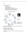
The eyeball can be considered to be a ball of three coats:
1. The outer, tough fibrous sclera forming the white of the eye and coming to the
front of the eye to form the curved transparent cornea. The cornea is
responsible for most refraction of the light.
2. The middle layer, the choroid contains many blood vessels and is darkly
pigmented to prevent the reflection of light inside the eye ensuring a sharp
image. The choroid forms the iris in the front of the eye. The iris can be blue,
brown, grey or grey in humans. This is a thin round curtain-like structure with
radial and circular muscles. The pupil is the central hole in this curtain and it
is its job to control the amount if light entering the eye.
In dim light the radial muscles of the iris contract to dilate the pupil. In bright
conditions, the circular muscles of the iris contract to constrict the pupil, to
reduce the amount of light entering.
3. The retina, which converts light into electrical impulses, is the inner coat and it
consists of light receptor cells called rods and cones. The 100 million rods
respond to light stimulus of black, white and shades of grey. The 7 million cones
respond to coloured light rays.
The cones are concentrated in an area called the fovea or yellow spot, which is
where the human focuses his images. It is part of the retina with the clearest
vision.
The rods are arranged around the periphery of the retina, which explains why
at night (when the cones aren’t working because they need high light intensity)
you see well out the corner of your eye.
The blind spot is an area of the retina, which has no rods or cones because it is
at this point that all sensory neurons (each one attached to a rod or cone)
converge and exit the eye forming the optic nerve. The neurilemma, Schwann
cells and myelin all protect the sensory neurons.
The eye is divided internally by the lens and ciliary body into an anterior chamber
filled with aqueous humor and a posterior chamber filled with a jelly-like vitreous
humor. The ciliary body consists of the ciliary muscle and the ciliary processes. The
ciliary muscle is donut shaped and has suspended from its internal surfaces the non-
elastic suspensory ligaments, which suspend the elastic, biconvex, transparent lens at
their center.
The eye:
Description of the eye:
The eye fits into and is protected by the bony eye orbit, at the back of which is a pad of
fat. The upper and lower eyelids with their eyelashes protect the eyeball.
The tear gland (lachrymal gland) is situated in the upper, outer corner and it secretes an
antiseptic fluid, which keeps the eyeball moist and washes away any foreign bodies.
Surplus tears drain into the nasal cavity.
The exposed surfaces of the eyeball and the inner surfaces of the eyelids are covered by a
delicate membrane – the conjunctiva.
The eyeball can be considered to be a ball of three coats:
1. The outer, tough fibrous sclera forming the white of the eye and coming to the
front of the eye to form the curved transparent cornea. The cornea is
responsible for most refraction of the light.
2. The middle layer, the choroid contains many blood vessels and is darkly
pigmented to prevent the reflection of light inside the eye ensuring a sharp
image. The choroid forms the iris in the front of the eye. The iris can be blue,
brown, grey or grey in humans. This is a thin round curtain-like structure with
radial and circular muscles. The pupil is the central hole in this curtain and it
is its job to control the amount if light entering the eye.
In dim light the radial muscles of the iris contract to dilate the pupil. In bright
conditions, the circular muscles of the iris contract to constrict the pupil, to
reduce the amount of light entering.
3. The retina, which converts light into electrical impulses, is the inner coat and it
consists of light receptor cells called rods and cones. The 100 million rods
respond to light stimulus of black, white and shades of grey. The 7 million cones
respond to coloured light rays.
The cones are concentrated in an area called the fovea or yellow spot, which is
where the human focuses his images. It is part of the retina with the clearest
vision.
The rods are arranged around the periphery of the retina, which explains why
at night (when the cones aren’t working because they need high light intensity)
you see well out the corner of your eye.
The blind spot is an area of the retina, which has no rods or cones because it is
at this point that all sensory neurons (each one attached to a rod or cone)
converge and exit the eye forming the optic nerve. The neurilemma, Schwann
cells and myelin all protect the sensory neurons.
The eye is divided internally by the lens and ciliary body into an anterior chamber
filled with aqueous humor and a posterior chamber filled with a jelly-like vitreous
humor. The ciliary body consists of the ciliary muscle and the ciliary processes. The
ciliary muscle is donut shaped and has suspended from its internal surfaces the non-
elastic suspensory ligaments, which suspend the elastic, biconvex, transparent lens at
their center.