IB SEHS unit 4 already rated A+
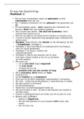
IB SEHS unit 4 already rated A+ label a motor unit - dendrite - cell body - nucleus - axon - motor end plate - synapse - muscle dendrites receives impulses from other neurons and pass them to the cell body Best Small School Prospects in the 2022 NFL Draft cell body processes the info from the dendrites and sends an impulse down the axon Axon acts as a wire, carrying the electrical impulse Myelin sheaths surround the axon, protecting it. They act as insulators, which speed up the impulse motor end plate connection between neuron and muscle synapse the junction between the dendrites of another neuron or where they meet the muscle fibre acetylcholine to contract muscles cholinesterase enzyme that breaks down acetylcholine (muscles relaxation) Explain how skeletal muscle contracts by the sliding filament theory 1. action potential arrives at the neuromuscular junction 2. cell membrane is depolarized, acetylcholine is released, sodium goes into cell 3. calcium is released from sarcoplasmic reticulum into muscle 4. calcium binds to troponin (on tropomyosin) causes movement and revealing myosin binding sites on actin 5. ATP is hydrolyzed to form ADP + phosphate 6. myosin head binds to actin and forms cross bridges (stays there until ATP molecule releases it) - if calcium is still there cross bridge is still there 7. ADP is released causing myosin heads to activate and move towards center of sarcomere (Power stroke) 8. power stroke is continued till z lines are pulled toward H zone 9. calcium is transported back causing termination of cross bridge. 10. myosin binding sites covered by tropomyosin and troponin (returns to original state) myofilament The structure that makes up the myofibril, examples include Actin and Myosin. sarcromere a section of muscle fibre I band thin filaments only (Actin) A band The band of the sarcomere that extends the full length of the thick filament. Does not change in length when muscle is contracted or relaxed. Z line end of sarcomere H zone thick filaments only (myosin) sarcoplasmic reticulum store of calcium flexion Decreasing the angle of a joint extension Increasing the angle at a joint rotation Movement around a pivotal point plantar flexion Pointing of the toes/ foot towards the body - increasing angle between tibia and foot dorsiflexion Raising of the foot/toes towards the body - Decreasing angle between tibia and foot
Written for
- Institution
- IB SEHS unit 4 already rated A+
- Course
- IB SEHS unit 4 already rated A+
Document information
- Uploaded on
- April 19, 2024
- Number of pages
- 9
- Written in
- 2023/2024
- Type
- Exam (elaborations)
- Contains
- Questions & answers
Subjects
-
ib sehs unit 4 already rated a
Document also available in package deal