Volledige uitwerking van de practica, met uitleg, afbeeldingen en antwoorden. MET oefenexamen met volledige uitleg hoe je dit aanpakt.
Handleiding van EEG is ook beschikbaar in dit format.
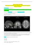
Practicum 5: Understand the structure of a 3D MRI scan, learn to navigate in a standard brain, work
with brain atlases
FSL openen via terminal XQuartz, FSLeyes openen via ‘fsleyes’ intypen in terminal.
“nii” = Nifti format; standaard voor MRI data.
“gz” = file is compressed
Nu kijken naar bestand “MNI152_T1_1mm.nii.gz” à Gemiddelde van 152 hersenen.
Do you expect more alignment (= less blurring) between different brains at central or at peripheral regions of
the brain? à Centrale regios zullen meer alignt zijn.
The 152 different brains have all been transformed into the same space. There is less variation near the
center of these brains than in the individual gyri and sulci. Therefore, there is more overlap (and less
blurring) between brains at central compared to peripheral regions.
Klik op (i) = algemene info, info over voxels, oriëntatie.
X-as = dim1
Y-as = dim2
Z-as = dim3
1 voxel = 1 mm x 1 mm x 1 mm = kubieke millimeter = 1 mm3
Een brein image is ongeveer 7 liter.
,“Storage order = Radiological” = rechter kant van het brein is aan de linker kant van het plaatje.
X = rechts
Y = front
Z = top
Coördinaten (0,0,0) = Centrum van afbeelding. Dichtbij de Anterior Commissure (AC).
Coördinaten (0,2,-4) = AC.
Due to the "nodding" of the head (i.e., chin on chest), the cerebellum is slightly more superior in this image.
Due to the "nodding" of the head (i.e., chin on chest), the posterior end of the corpus callosum is slightly
more superior in this image.
The anterior end of the corpus callosum is at about y = +32 mm, the posterior end is at about y = -43 mm.
We have to add the 1 mm slice right in the middle at y = 0, which leads to a length of 32+43+1 = 76 mm.
“Ga naar coördinaat (…) Welke structuur is dit?” à Benoem Rechts of Links!
, Atlas informatie: Settings à OrthoView1 à Atlas panel
Note that this is a probabilistic atlas: the further removed from the center of a region, the smaller the
probability that a voxel is part of this region, and the darker the color. The white colors in the center have a
very high probability of belonging to this structure.
As the names imply, the Harvard-Oxford Cortical Atlas contains cortical grey matter structures, and the
Harvard-Oxford Subcortical atlas contains subcortical grey matter structures.
Voordelen van het kopen van samenvattingen bij Stuvia op een rij:
Verzekerd van kwaliteit door reviews
Stuvia-klanten hebben meer dan 700.000 samenvattingen beoordeeld. Zo weet je zeker dat je de beste documenten koopt!
Snel en makkelijk kopen
Je betaalt supersnel en eenmalig met iDeal, creditcard of Stuvia-tegoed voor de samenvatting. Zonder lidmaatschap.
Focus op de essentie
Samenvattingen worden geschreven voor en door anderen. Daarom zijn de samenvattingen altijd betrouwbaar en actueel. Zo kom je snel tot de kern!
Veelgestelde vragen
Wat krijg ik als ik dit document koop?
Je krijgt een PDF, die direct beschikbaar is na je aankoop. Het gekochte document is altijd, overal en oneindig toegankelijk via je profiel.
Tevredenheidsgarantie: hoe werkt dat?
Onze tevredenheidsgarantie zorgt ervoor dat je altijd een studiedocument vindt dat goed bij je past. Je vult een formulier in en onze klantenservice regelt de rest.
Van wie koop ik deze samenvatting?
Stuvia is een marktplaats, je koop dit document dus niet van ons, maar van verkoper stansterk. Stuvia faciliteert de betaling aan de verkoper.
Zit ik meteen vast aan een abonnement?
Nee, je koopt alleen deze samenvatting voor €5,29. Je zit daarna nergens aan vast.