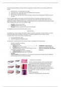
There are about 200 different cell types which are organized in tissues. These tissues can be classified into 4
groups:
1. Epithelial tissue – the outer layer of an organ.
2. Connective tissue – layer that connect the epithelial and muscle layer.
3. Muscle tissue – the layer that moves.
4. Nervous tissue – the layer that controls the body’s movement by sending signals to different parts of
the body.
These four types together form organs, such as the gut. Blood is an example of connective tissue, since it
connect different cell types and all cells from the connective tissue share the same origin, which is the case
with blood in embryo’s. With embryonic origin, cell types that are part from the same basic tissue type can look
different! What these cells share is the embryonic origin.
- Ectoderm: nervous tissue → skin.
- Endotherm: digestive tissue → lung
- Mesoderm: muscle, connective tissue (from mesenchyme).
The epithelial tissue can be derived from all three classes.
Epithelial tissue
An epithelium is a tissue existing of cells that are tightly connected to each other. It is derived from one of the
three primary germ layers (ectoderm, endotherm or mesoderm). Epithelial tissue is always on top of
connective tissue. There are two types of epithelial tissue:
1. Covering epithelia (e.g. skin),
2. Glands (invagination of epithelial layers)
An epithelium is classified based on its embryonic origin, morphology and cell organization:
- Ectoderm, endoderm, mesoderm,
- The shape of the cells
I. Squamous (flat cells) Transitional: changed between
II. Cuboidal (squares, cubic-shaped) squamous and cuboidal. When you
III. Columnar (elongated, often has small structures on top) stretch out cuboidal cells, they become
- The number of cell layers (strata) squamous.
I. Simple epithelium (one layer of epithelia),
II. Stratified epithelium (more layers of epithelia)
- Stratified,
- Pseudo-stratified (looks stratified, but is not, because the nuclei of the epithelial cells are at different
levels leading to the illusion of being stratified).
characteristics of epithelia:
- There is no blood circulation as well as no blood vessels,
- They are polarized (apical, basal side and lateral side).
Apical: the microvilli, the small structures on top of the
epithelia. Basal side: the side that faces the basement
membrane, like the connective tissue layer the cell lives on
(the bottom edge of the cell). Lateral side: the outer side of
the cell (zijkanten, de zijden die de cellen met elkaar
verbindt)
- Surface specializations,
- Presence of a basal membrane on the basal side.
, Apical domain:
- Microvilli: increases the surface area (cell surface). It helps the
adsorption of compounds, the plasma membrane is larger due to the
microvilli. This helps the cell to have more contact with exocellular
environment to take up compounds from the liquid on top of the
cell. The microvilli (short fingers) contains cytoskeleton of actin
(microfilaments)
- Cilia: movement (they can be used to move the liquid on top of the
cell), sensory function. The cilia contains cytoskeleton of microtubuli
(tubulin).
There is an extension of the cytoskeleton and the plasma The cytoskeleton of cilia is 5x larger than
microvilli. They membrane folds around the extended cytoskeleton,
which are narrow and long. The cytoskeleton is made from
increases the interaction with the extracellular environment from microtubuli. The cilia can move
because of the to adsorb compounds. The cytoskeleton is
made from actin! Contractions from the microtubuli inside the cilia.
Tight junction
Tight junction
Also called the Zonula Occludens. It prevents small
molecules to come inside the epithelial cells. The
Adhesion cell membrane of one cell is connected to the cell
belt membrane of the adjacent cell via the tight junction
Terminal proteins. these tight junction proteins function as a
web ‘zipper’ and the tight junction is around the whole
cell!
Button
desmosome
Functions:
- It prevents the transport between cells,
- Membrane proteins of both cells are
compartmentalized (apical and basolateral) to keep
them separated. Membrane proteins cannot go
from the top of the cell (apical side) to the bottom
of the cell (basolateral side) due to the tight
junction.
Hemidesmosome
(a)
Adhesion belt
Gap junctions
Also called the Zonula adhearens. It is also around
, the cell and it is made by transmembrane-linker proteins called cadherins. The plasma membrane is widened
(intercellular) and these adhesion belt molecules are attached to actin (intracellular). The adhesion belt
connects two cells by the interaction of actin and cadherin.
Gap junction
It is also called nexus. Gap junctions are pores in the membranes by which the adjacent cells can communicate
with each other by passing ions, amino acids, molecules, hormones and electrical impulses. It allows
Intercellular transport: 1.5 nm. The proteins that make up the gap junctions are called the connexins.
Button desmosome and hemidesmosome
Button-like spots found all around the cell that bind adjacent cells together. They are not around the whole cell
and it makes the strongest connection between cells because of the intermediate filaments (which are thicker
than actin, which leads to a stronger connection). Examples of intermediate filaments are keratin, vimentin,
desmin etc.) There are also button-like spots that bind the cell to the tissue underneath (connective tissue)
which are called the hemidesmosome. Hemidesmosomes bind to fibers (called basal lamina) outside the cell.
The basal lamina consists of two layers: Lamina Lucida (lamina that is light, has few fibers) and Lamina densa
(lamina that is dense, meaning it has more fibers). The epithelial cell uses adhesion molecules to bind to these
fibers in the basal lamina. The connective tissue is also bound to the basal lamina, so therefore, the basal
lamina acts like a ‘glue’ that binds the epithelial cell to the connective tissue layer.
Basal domain
The basal domain consists of the hemidesmosomes, basal lamina and plasma membrane invaginations
(infolding of the basal membrane which causes basal labyrinth). This is needed to increase the surface area.
The basal labyrinth is like the microvilli (fingers). Due to the basal labyrinth, there is a lot of plasma membrane
to take up oxygen and glucose from the blood. The dark structures are the mitochondria due to the fact that
oxygen and glucose come in. These two need to be converted into energy.


