Histology year 1
The cell is the essential building block of life on earth. It has a limiting membrane (inside/outside compartments). It
contains biomolecules (e.g. protein/RNA/DNA). It is an autonomous unit in performing a function. It can respond and adapt
to stimuli and it can often reproduce itself. An exception is the neuron and red blood cell, they cannot divide by itself
obviously. In 1665, the cells were first discovered by Robert Hooke. He was also involved in the development of the
microscopes. Cells can be diverse. despite the obvious differences between cells, basic functions and components are highly
similar.
The form of the protein is responsible for its function. From
proteins to cell behaviour: organelles. Bacteria has no nucleus or
other organelles, Archaea has no nucleus but often extremophiles
(being able to live under extreme conditions), eukaryotes have a
nucleus and organelles (sometimes multicellular life forms). The
microscope is invented by Antonie van Leeuwenhoek and is
essential to study cells. Antonie was good in making lenses.
- Condenser lens: allows the light coming out lamp to direct it into
a narrow beam that is projected onto the sample.
- Objective lens: lens where you can change the magnification and
distances from.
An advanced form of light electroscope: fluorescent microscopy.
,Histology year 1
A type of light reaches the beam splitting mirror and gets directed onto the object via the objective lens. The
object contains a fluorescent protein, meaning that that protein can accept light of a certain wavelength (so a certain
colour) and after that they fall back in their energy state in which they emit light over a longer waveform. Jelly fish are
transparent in live in a transparent environment. How do they attract others to mate? → they have bioluminescence that
can send out a specific type of light (green fluorescent light). They have molecules that send out light. Using GFP, you can
visualise subpopulations of cells in tissue. By putting the tissue under the microscope, you can see where the GFP lights up.
You can see the function of different cells in the tissue just by looking where the GFP lights up, meaning the areas where
the proteins are being expressed (gene is thus active). GFP can also be linked to certain molecules that function within cells.
Using GFP, you can visualise living cells in multicolour, due to the fact that fluorescence has multiple colours. Green light
has a wavelength of 500 nm.
Electron microscope
With the EM, you can zoom more precise on the objects but under one condition: it must be dead tissue, whereas in LM
you can zoom in on live and dead objects. You can zoom more with the EM because you use electrons instead of photons.
Electrons have a wavelength that is 1000 times smaller than the wavelength of photons, allowing us to zoom into details.
Lights have different wavelengths and the smaller the wavelength is (EM), the more detailed and zoomed in it will be. If the
wavelength is large, than you would not be able to zoom into details.
The EM is under high vacuum, because the electrons are easily scattered by small dust particles and if you can see a single
molecule, this molecule can disturb the visuals. Also, we do not use glass lenses; instead, we use electromagnetic lenses.
,Histology year 1
Protons from the atoms in the objects attract the electrons from the microscope, resulting in the bending of the electrons
in the preferred way. In EM, heavy metals are used that contain a lot of protons which can diverge the electrons away and
thereby we can see something.
Advantages of EM compared to LM:
- Better resolution (up to 0,5 nm, atoms)
- You are able to visualise the whole cell, not only a fluorescent probe (like GFP)
- There is a huge magnification range
Disadvantages of EM compared to LM:
- Requires fixation of cells (operates under vacuum), all the cells contain water, and if you put that cell directly
under the EM, the water would evaporate immediately, meaning that the whole cell would collapse.
- Only small pieces of tissues can be imaged, the amount of tissue that we can examine at the same time is limited.
- Time-consuming method
- We cannot look at living cells.
All life on Earth consists of cells. A cell is a compartment containing biomolecules (DNA, RNA, protein) that are surrounded
by membrane and can react to stimuli. All cells have the same basic ingredients but the show a huge diversity in shapes and
specialised functions. To study the form-function relationships, microscopes are essential: Light microscopy, Fluorescence
microscopy, Electron microscopy . Using these methods in combination, you can study processes in cells at a 1 000 000
magnification range.
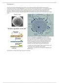
Nucleus
Here, the genetic material (DNA) is stored of an
eukaryote. The nucleus has a shape that is related to
its function. The nucleus also has an envelope
surround it called the nuclear envelope. Inside the
nucleus, you see dark and light spots which are the
loose and condensed chromatin. The nuclear
envelope distinguishes the genetic material from the
rest of the cell. The nuclear envelope not only keeps
the DNA in, but also keeps selected molecules out
that could damage the DNA. Transcription happens in
the nucleolus and via the nuclear pore, the
transcribed RNA goes to the ribosomes on the ER and
gets translated into amino acids and proteins. The
nucleolus is dense, because there is a high
concentration of protein here. You need many
proteins for transcription.
- Chromatin: the DNA-protein complex that forms chromosomes.
, Histology year 1
Translation of RNA to protein happens in the rough endoplasmic reticulum.
In most cells, the ER is found everywhere. The ER is important to make
luminal proteins.
Golgi
apparatus
Responsible
for protein
modification
and protein
sorting. It
adds or removes sugar groups from the to proteins.
Mitochondrion
Produces ATP (energy). Electro transporting molecules
are here which are membrane based proteins. Burning
of glucose yields energy in the form of adenosine
triphosphate (ATP). Mitochondria also contain their
own DNA. They originate from bacteria. Chloroplasts
are also derived from symbiotic bacteria. The cell went
into a symbiotic relationship with this bacterium,
resulting in the origin of chloroplast (the same for
mitochondrion). Chloroplasts are responsible for the
energy supply in cells via photosynthesis. However,
plants need chloroplasts and mitochondrion in order to
burn glucose.
Cytoplasm (soup)
The liquid in which the organelles swim inside the cell.
Cytoplasm is not a fluid, but a jelly matrix of proteins. Processes that take place in the cytoplasm are: transport, protein
synthesis, protein break-down, signal transduction, membrane fusion, ionic homeostasis.
cytoskeleton
Special group of molecules that organize into
filaments that support the shape of the cell and
they make sure that organelles are able to take up
a certain place in the cell in respective to each
other. Microtubule is for the shape of the cell,
actin for the mobility of the shape and
intermediate filaments link everything together
and are important for connections between other
cells in a multicellular organism.
Cells in the tree of life comprise bacteria, archaea
and eukaryotes. Only eukaryotes contain
organelles and a nucleus, these cells can form
multicellular organisms. • The basic organelles and their function: The nucleus: store DNA and transcription of DNA to RNA.
The endoplasmic reticulum: translation of RNA to protein and protein folding. Golgi apparatus: post-translational


