100% tevredenheidsgarantie Direct beschikbaar na betaling Zowel online als in PDF Je zit nergens aan vast
logo-home
Very extensive summary of the lectures €2,99
In winkelwagen

Samenvatting

Very extensive summary of the lectures

 112 keer bekeken  5 keer verkocht

So after listening to all the lectures and taking almost constant notes, I can guarantee you these pages will help (if you can get through my mental notes here and there). I know for a fact that this is one of the longest summaries for this course and it will be worth it. NOTE: lecture order is mes...

[Meer zien]
Voorbeeld 4 van de 69  pagina's

  • 10 juli 2019
  • 69
  • 2018/2019
  • Samenvatting
Alle documenten voor dit vak (19)
avatar-seller
juliaelisadrijver
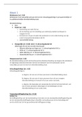
Lecture 1: The Adolescent Brain


 Video about what adolescents think about how the brain
works.

-> they're very visual, but they don't seem to have thought many
things through. They don't really think about the perspectives of
others well


 Bring adolescent development into a neuroscientific
perspective, bridging neural and social emotional and
cognitive development.
 Use neuroscientific theories to explain adolescent
development.

QUIZ
Q1. The following methods were the first that studied the brain,
and were established in the 19th century. Which methods yielded
valuable knowledge about the brain that is still valid/being used?

1. Post-mortem studies
2. Phrenology
3. Lesion studies


Why not phrenology? It's a localization theory that is inaccurate,
developed by poor mr. Gall. It's important to realise that he did
bring about the idea of localized function theory.
Post-mortem studies: This is how Broca and Wernicke found their
areas.
Lesion studies: Think of Phineas Gage. Medial prefrontal cortex
and orbitofrontal cortex damage leads to a total personality
gage.
How do we study the brain


Q2. The brain consists of several anatomically distinguishable
lobes. In the picture we can see the:

, 1. Frontal Lobe
2. Occipital Lobe
3. Temporal Lobe
4. Parietal lobe
5. Medial Lobe
6. Cerebellum


Q3. The brain consists of gyri and sulci to increase the surface of
the cortex. Which is which in this picture?


1. 1= gyrus, 2= sulcus
2. 1= sulcus 2= gyrus


Q4. For each of the following statements, indicate whether it
applies to white matter (WM) or gray matter (GM) in the brain

 Connections between brain areas
 Consists of neuronal cell bodies
 Surface of cerebral cortex & cerebellum
 Is myelinated
 In subcortical structures

, 1. WM = 1+2+4; GM = 3+5
2. WM = 1+4 ; GM = 2+3+5
3. WM = 2+3+5 ; GM = 1+4
4. WM = 1+4+5; GM = 2+3


Q5. Which part of the neuron enables enhanced/optimal
neurotransmission?


1. Axon
2. Synpase
3. Glia
4. Myoline
5. Neutrostransmitter
6. Myelin
7. Dendrite
8. Synapse
9. Soma


Q6. Which part of a neuron conducts electrical impulses?

1. Axon
2. Synpase
3. Glia
4. Myoline
5. Neutrostransmitter
6. Myelin
7. Dendrite
8. Synapse
9. Soma


Q7. With age, there are changes in brain function, but no changes
in brain structure

1. Yes
2. No


Q8. Who has more brain cells?

, 1. An 11 year old
2. An 80 year old


Q9. What happens with grey matter during adolescence?

1. It increases
2. It decreases


By the age of 8, the brain size is about 80% of the adult size. One
of the biggest changes that happen in the brain of growing
children are the increasing amount of connections between brain
cells, and there is an increase in myelination of the cortex.
Therefor, white matter increases. Gray matter decreases though.
First it increases, then it decreases. Why is this decrease not
linear? Well there is synaptogenesis, an increase in synapses. But
there's also pruning, where excess synapses are eliminated.

Q10. What is the pattern of structural change in the brain

1. From back to front
2. From front to back
3. Currently unknown


The amount of gray matter decreases in the more back areas (so
occipital lobe ->parietal lobe -> frontal lobe) first, and only then
starts to decrease in the more frontal areas.

Q11. FMRI technique on which of the following signals?


1. EEG (Electroencephalography)
2. REM (Rapid eye Movement)
3. BOLD( Blood-Oxygen Level dependent)
4. MEG ( Magnetoencephalography )


MRI - Magnetic resonance imaging makes images of the brain
structure
FMRI - functional, shows activity.
Indirect measure of brain activity. As soon as you use an area, it
will require blood, usually oxygenated. There will be changing

Voordelen van het kopen van samenvattingen bij Stuvia op een rij:

Verzekerd van kwaliteit door reviews

Verzekerd van kwaliteit door reviews

Stuvia-klanten hebben meer dan 700.000 samenvattingen beoordeeld. Zo weet je zeker dat je de beste documenten koopt!

Snel en makkelijk kopen

Snel en makkelijk kopen

Je betaalt supersnel en eenmalig met iDeal, creditcard of Stuvia-tegoed voor de samenvatting. Zonder lidmaatschap.

Focus op de essentie

Focus op de essentie

Samenvattingen worden geschreven voor en door anderen. Daarom zijn de samenvattingen altijd betrouwbaar en actueel. Zo kom je snel tot de kern!

Veelgestelde vragen

Wat krijg ik als ik dit document koop?

Je krijgt een PDF, die direct beschikbaar is na je aankoop. Het gekochte document is altijd, overal en oneindig toegankelijk via je profiel.

Tevredenheidsgarantie: hoe werkt dat?

Onze tevredenheidsgarantie zorgt ervoor dat je altijd een studiedocument vindt dat goed bij je past. Je vult een formulier in en onze klantenservice regelt de rest.

Van wie koop ik deze samenvatting?

Stuvia is een marktplaats, je koop dit document dus niet van ons, maar van verkoper juliaelisadrijver. Stuvia faciliteert de betaling aan de verkoper.

Zit ik meteen vast aan een abonnement?

Nee, je koopt alleen deze samenvatting voor €2,99. Je zit daarna nergens aan vast.

Is Stuvia te vertrouwen?

4,6 sterren op Google & Trustpilot (+1000 reviews)

Afgelopen 30 dagen zijn er 51683 samenvattingen verkocht

Opgericht in 2010, al 15 jaar dé plek om samenvattingen te kopen

Start met verkopen
€2,99  5x  verkocht
  • (0)
In winkelwagen
Toegevoegd