Case 7 hunger and thirst
1. What is hunger?
- the system for controlling food intake and energy
balance is significantly more complex than those
controlling thermoregulation and fluid balance. One
important reason for this greater complexity is that we
need food to supply not only energy, but also crucial
nutrients. the nervous system not only monitors
nutrient and energy levels and controls digestion (the
process of breaking down ingested food), but also has
complex mechanisms for anticipating future
requirements.
- Alloestaesis: coordinated set of behavioural and
physiological changes to maintain homeostasis
- The cephalic phase is the preparatory phase; it often
begins with the sight, smell, or even just the thought of
food, and it ends when the food starts to be absorbed into the bloodstream. The
absorptive phase is the period during which the energy absorbed into the bloodstream
from the meal is meeting the body’s immediate energy needs. The fasting phase is the
period during which all of the unstored energy from the previous meal has been used
and the body is withdrawing energy from its reserves to meet its immediate energy
requirements; it ends with the beginning of the next cephalic phase
- basal metabolism: processes such as heat production, maintenance of membrane
potentials, and all the other basic life-sustaining functions of the body.
- Because people and animals adjust their metabolism in response to under- or
overnutrition, they tend to resist either losing or gaining weight
- For short-term storage, glucose can be converted into a more complicated molecule
called glycogen and stored as reserve fuel in several locations, most notably the liver
and skeletal muscles. This process, called glycogenesis, is promoted by the pancreatic
hormone insulin. A second pancreatic hormone, glucagon, mediates the conversion of
glycogen back into glucose, a process known as glycogenolysis, which is triggered
when blood concentrations of glucose drop too low.
- For longer-term storage, fats are deposited in the fat-storing cells that form adipose
tissue.
- Another important role of insulin is enabling the body to use glucose. Most cells
regulate the import of glucose molecules via glucose transporters: specialized proteins
that span the cell membrane and bring glucose molecules from outside the cell into the
cell for use. The glucose transporters must interact with insulin in order to function.
- Release insulin
o 1. Cathalic phase: The sensory stimuli from food (sight, smell, and taste) evoke
a conditioned release of insulin in anticipation of glucose arrival in the blood.
This release, because it is mediated by the brain, is called the cephalic phase of
insulin release (recall that cephalon means “head”).
o 2. During the digestive phase, food entering the stomach and intestines causes
them to release gut hormones, some of which stimulate the pancreas to release
insulin The digestive tract contains taste receptors like those found on the
tongue; these provide further regulation of insulin release
1
, o 3. During the absorptive phase, special cells in the liver, called glucodetectors,
detect the glucose entering the bloodstream and signal the pancreas to release
insulin.
- Type 1 diabetes: no production insulin, type 2 diabetes: insensitivity to insulin
- The newly released insulin enables the body to make use of some of the glucose
immediately and prompts the conversion of extra glucose into glycogen, which is then
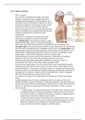
stored in the liver and muscles. The liver communicates with the pancreas via the
nervous system. Information from glucodetectors in the liver travels via the vagus
nerve to the nucleus of the solitary tract (NST) in the brainstem and is relayed to the
hypothalamus. Efferent fibers carry signals from the brainstem back out the vagus
nerve to the pancreas. These efferent fibers modulate insulin release from the
pancreas.
- low levels of insulin between meals could signal the brain to make us feel hunger,
impelling us to find food and eat. insulin can provide a satiety signal to the brain.
Somehow the brain integrates insulin and glucose levels with other sources of
information to decide whether to initiate eating
- ventromedial hypothalamus: satiety centre
- lateral hypothalamus: hunger centre
- arcuate nucleus of the hypothalamus contains a highly specialized appetite controller
that is governed by circulating levels of a variety of hormones
- leptin: the brain seems to monitor circulating leptin levels to measure and regulate the
body’s energy reserves in the form of fat. Defects in leptin production or leptin
sensitivity cause a false underreporting of body fat and lead to overeating, especially
high-fat or sugary foods.
- Ghrelin: appetite stimulant, Circulating levels of ghrelin rise during fasting and
immediately drop after a meal is eaten, stomach releases it in bloodstream.
- PYY3-36: may act in opposition to ghrelin, providing a potent appetite-suppressing
stimulus to the hypothalamus. Released by intestants.
- Cck: released by small intestants, can increase satiety, CCK, induces illness: CCK
administered to rats after they have eaten an unfamiliar substance induces a
conditioned taste aversion for that substance, and CCK induces nausea in humans
- Arcuate appetite system: The appetite system relies on two sets of arcuate neurons
with opposing effects, which are named according to the types of neurotransmitters
they produce. When activated, arcuate neurons that produce the peptides pro-
opiomelanocortin (POMC) and cocaine- and amphetamine-regulated transcript
(CART)—called POMC neurons for short—act as satiety neurons, inhibiting appetite
and increasing metabolism. In contrast, the other set of neurons—called NPY neurons
because they produce neuropeptide Y (NPY) along with agouti-related peptide
(AgRP)—act as hunger neurons when activated, stimulating appetite directly,
inhibiting POMC neurons (thereby blocking satiety signals), and reducing metabolism,
a set of actions that promotes eating and weight gain. Projections from the POMC
neurons and NPY neurons also leave the arcuate and make contact with neurons in
other hypothalamic sites. It is through these projections that the arcuate system
ultimately modulates food intake. High circulating levels of leptin activate the
appetite-suppressing POMC neurons but inhibit the appetite-increasing NPY neurons,
so in both systems leptin is working to suppress hunger. In contrast to leptin, ghrelin
and PYY3-36 provide more-rapid, hour-to-hour hunger signals from the stomach and
gut. Both peptides act primarily on the appetite stimulating NPY neurons of the
arcuate. Ghrelin stimulates these cells, leading to a corresponding increase in appetite.
PYY3-36 works in opposition, inhibiting the same NPY cells to reduce appetite.
2