Module 2: Tumor Immunology (Week 1)
LECTURE 1: BASIC TUMOR IMMUNOLOGY Monday 1/10/2018
1. Lymphoid-Myeloid differentiation patterns
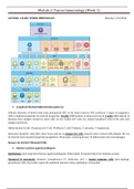
Cellular elements of blood arise from pluripotent HSC in the bone marrow. HSC produces 2 types of progenitor
cells: lymphoid progenitor & myeloid progenitor. B cells (differentiate in bone marrow) & T cells (differentiate in
thymus) have antigen receptors; other cells do not. Unlike B/T cells, ILC (innate lymphoid cells) & NK cells lack
antigen specificity.
Differentiated B cells plasma cell, T cell effector T cell (T helpers, T cytotoxic, T regulatory).
Immature dendritic cells (DC): enter tissue and act as phagocytic cells, matures after contact with antigen; DC can
be derived from myeloid/lymphoid progenitors. Monocytes: entering tissues differentiate into macrophages
Mature DC DO NOT PHAGOCYTISE
2. Defense systems against pathogens
Epithelium: most important barrier against pathogens, if disrupted require help from immune system
Chemical & enzymatic elements (complement C3, defensins, etc) + innate immune cells (macrophage,
granulocyte, NK) provide rapid cell-mediated response when epithelium is breached
, Module 2: Tumor Immunology (Week 1)
Adaptive immune response slower process, able to induce memory to provide faster & more efficient
response on 2nd exposure to the same antigen
3. Adaptive response (antibody-mediated)
Lag phase: body produces antibodies pathogens are being neutralized by cellular response.
Primary response: immunoglobulin level increases in serum (decreases when infection is overcome).
Secondary response: quicker & higher effectivity on 2nd encounter (memory cells involvement don’t need
much activation).
4. Antigen-presenting cells (APC) – key players in immune system
2 professional APC: dendritic cells & macrophages taking up antigens to break them down (into oligopeptides)
& present them to MHC class I/II. DC: watchdogs of the body, always patrolling for abnormal cells, Macrophages:
scavengers
DC maturation: precursors in bone marrow (marker: CD34+) exposed to cytokines (GM-CSF, IL-4)
immature DC (in resting/non-infected tissue, can only take up antigen, marker: CD1a+) exposure to antigen OR
presence of other cytokines (TNF alpha, CD40, LPS, GM-CSF, IL-4) mature DC (able to take up antigen & present
antigen to MHC, marker: CD83+)
, Module 2: Tumor Immunology (Week 1)
Tumor antigen processing by APC:
Tumor starts growing, some dies d/t necrosis release tumor
antigens tumor antigens recognized by DC taken up by DC
& into lymph nodes exposure of DC & tumor antigen to naïve T
cells T cell maturation (bound to DC) activation &
proliferation of T cells T cells migrate to tumor site
eradication by apoptosis
5. Mellman cancer immunity cycle
Destruction of tumor cells by chemotherapy,
radiation, or targeted therapy releasing
TAA/TSA TAA/TSA taken up by APC (uptake
can be enhanced by vaccine, IFN alfa, GM-CSF,
CD40 agonist, or TLR agonist) antigen
presentation priming & activation of naïve T
cells (enhanced by anti-CTLA4, IL-2, IL-12, etc)
activated T cell migrates into tumor
microenvironment infiltrate TME recognize
cancer cells (enhanced by CAR) killing of tumor
cells (enhanced by anti-PD1/PD-L1, IDO inhibitor)
, Module 2: Tumor Immunology (Week 1)
6. DC activity
DC receptors:
a. Fc receptor: binds to antigen-antibody
complex internalization
b. Toll-like receptors (TLR): binds viral &
bacterial antigens, can also bind to certain
ligands
Type I/II TLR: bacteria
Type III TLR: viral dsDNA (specific)
Type IV TLR: endotoxin/LPS
Type IX TLR: CpG hypomethylation
(virus/bacteria)
c. CD36 receptor: binds apoptotic bodies
d. CD91 receptor: binds heat shock proteins
Immature DC is activated via its TLR when presented to microbes receptor ligands can also bind to TLR to
mimic actual bacterial infection (i.e. vaccination strategy)


