Samenvatting
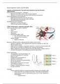
Summary Neurocognition week 1: Development: The brain and cognition over the life span
- Vak
- Neurocognition
- Instelling
- Universiteit Leiden (UL)
This document consists of extensive notes of the lecture of week 1 and the seminar and a summary of the article and chapter 5 and 15 of the book.
[Meer zien]