This summary is everything you will need for the tutorials and the exam. You will find all the mandatory reading for course 2.4 Perception summarized by an Honours student.
I like to create helpful visuals, overviews, and diagrams accompanied by explanations. I hope you'll enjoy them, and good luc...
2.4 Perception
Problem 1 – The Eye
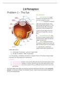
The 3 Membranes
1. Sclera/Fibrous tunic: tough,
protective covering (the white of our
eye) with transparent cornea
a. Cornea: transparent membrane
Light enters passing through cornea
which sharply refracts/bends it → The
refraction focuses light on the retina
(does most of the focusing process)
Is rigid and cannot adjust how much
light passes through (this is done by
lens)
2. Choroid/Vascular tunic: lines
the interior of sclera, contains most of
the blood vessels (supply the eye with
oxygen and nutrients)
3. Retina: made up of neurons
including the receptors that convert the
light entering the eye into neural signals
Iris: coloured part, a small donut-
shaped muscle with an opening in the
middle (light enters)
Pupil: the opening – controlled by contraction or relaxation of the iris → controls intensity of light
entering the eye. Both pupils work simultaneously
3 Chambers
1. Anterior chamber: between cornea and iris, filled with clear thin fluid called aqueous humor
2. Posterior chamber: between iris and lens, filled with clear thin fluid called aqueous humor
3. Vitreous chamber: main interior portion of the eye, filled with vitreous humor a clear more
gel-like fluid
Both fluids slightly refract light, but the amount cannot be adjusted (just like with cornea). Intraocular
pressure – the pressure of fluids in the chambers must be > than air pressure (to prevent collapsing of
the eyes like deflated balls)
,Lens and Accommodation
Focal length: the distance from the lens at which the image of an object is
in focus when it is far away from the lens aka “optical infinity” = light rays
parallel to one another
• Weak lenses → don’t refract/bend the light much, relatively thin
and flat → long focal length
• Strong lenses → refract light sharply, relatively thick and rounded
→ short focal length
• Power of a lens = diopters = 1/(focal length) in meters
o e.g. focal length of 0.5m has a power of 2.0 diopters
(1/0.5=2)
• The mammalian eye focuses by adjusting the shape of the lens to change its focal length
o Edges of the lens are strtched by zonule fibers that conect the lens to choroid
o Ciliary muscles are also attached to choroid
▪ When relaxed → choroid can pull on zonule fibers → stretches the lens and
makes it relatively thin and flat → weak lens → long focal length (focusing on
distant objects)
▪ When contract → oppose the pull by the choroid on the zonule fibers → lens
not stretched as much → thicker, rounded shape → stronger lens with a shorter
focal length (focusing on near
objects)
Accommodation: this adjustment of the shape of lens
to focus on objects at different distances
Retina
Retinal image: a clear image on the retina of the optic
array
Anatomy of the retina:
Photoreceptors: transduce light into neural signals
• Rods: provide black and white vision in dim light
• Cones: provide high-acuity colour vision in bright light
o S-cones: most sensitive to short wavelengths of light
o M-cones: most sensitive to medium wavelengths of light
o L-cones: most sensitive to longer wavelengths of light
Axons of retinal ganglion cells (RGC) exit the eye in the optic disk/blind spot forming a bundle called
optic nerve.
,Fovea: where the optic axis passes through at the centre of retina
• Anatomy of fovea different from the rest of retina – No rods ONLY CONES
• Cones in fovea are thinner than elsewhere so they can be densely packed together (in a
hexagonal grid)
, • Ganglion cells and inner nuclear layers are pushed to the side of fovea → light reaches foveal
cones without being scattered as much → high-acuity vision at the centre of gaze
How does it work in the retina?
• Incoming light passes through other layers of neurons in the retina and strikes the outer parts
of the photoreceptors where it is transduced into neural signals expressed as changes in the
membrane potential of the photoreceptors
• The changes in photoreceptor membrane potential alter the amount of neurotransmitter
molecules that the photoreceptors release
• “Through pathway”: photoreceptors → bipolar cells → retinal ganglion cells
o The flow of neurotransmitter molecules released by rods and cones → affects the
membrane potential of bipolar cells → changes their release of neurotransmitter
molecules.
o The change in bipolar cell neurotransmitter release affects the membrane potential of
RGCs, which in turn affects their firing rates
• “Lateral pathway”: horizontal & amacrine cells
o Allows the presence of light at one location on the retina to affect the responses of
photoreceptors, bipolar cells, and RGCs at adjacent locations on the retina
o these lateral influences provide a way for the neural signals to transmit information
about luminance contrast (crucial role in detecting edges and boundaries)
• Retinal ganglion cells send action potentials to the brain via the optic nerve
GOAL OF EYE’S OPTICAL SYSTEM
1. Constriction/Dilation of the pupil by the iris → to control the amount of light entering the eye
2. Accommodation by the lens → focus the light on the retina
, 3. Form a clear image on the retina of the optic array → transformed into neural signals to be sent
to the brain
Light
• Electromagnetic spectrum -
produced by electric charges
and is radiated as waves
• Energy – described by its
wavelength – distance
between the peaks of the
electromagnetic waves
• Visible light - the energy
within the electromagnetic
spectrum that humans can
perceive, has wavelengths ranging from about 400 to 700 nanometers (nm)
• Apart from wavelength, light can be described in terms of photons – the smallest passible
packet of energy
The Eye
• Light reflected from objects in the environment
• Enters the eye through the pupil and is focused by the
cornea and lens to form sharp images of the objects on the
retina, which contains the receptors for vision
• Visual receptors → rods and cones
o Contain light-sensitive chemicals called pigments
that react to light → trigger electrical signals
o These signals flow through the network of neurons
in the retina → to the optic nerve → signals toward
the brain
Focusing by the eye
• Cornea – transparent covering of the eye → 80%, but fixed
in place so it cannot adjust its focus
• Lens – 20% of eye’s focusing power, can adjust (ciliary
muscles and zonule fibers) its shape to focus on stimuli
located in different distances
• Accommodation: ciliary muscles at the front of the eye
tighten and increase the curvature of the lens so that it gets
thicker → the curvature bends the light rays, which pull the focus point back to retina (image
becomes sharp)
• Near point: limit of accommodation → distance at which your lens can no longer adjust to bring
close objects into focus
, o The distance of the near point increases as a person gets older – presbyopia “old eye”
(20 y.o. around 10cm, 40 y.o. 22cm, 60 y.o. 100cm)
o The loss of ability to accommodate occurs because the lens hardens with age and ciliary
muscles become weaker → more difficult for the lens to change shape for vision in
closer range
Eyesight problems
Myopia/Near-sightedness: inability to see distant objects clearly
• Myopic eye brings the parallel rays of light into focus in front of the
retina → image reaching the retina is blurry
o Refractive myopia – cornea and/or lens bends the light too
much
o Axial myopia – eyeball is too long
• light into focus behind the retina → image reaching the retina is
blurry
Correction methods:
• Eyeglasses/corrective lenses – bending the incoming light so that it
reaches retina
• Laser-assisted in situ keratomileusis (LASIK) surgery – sculpting the
cornea so that it focuses light onto the retina
Transforming Light into Electricity
Transduction: light→electricity
• Conducted by rods and cones
• Key part – the outer segment –
containing a stack of discs
• Each disc contains thousands of visual
pigment molecules
o This molecule is a long strand of
protein called opsin which loops
back and forth 7 times
o Each visual pigment molecule
contains only one retinal
molecule – part of the visual
pigment that is sensitive to light
• Transduction is triggered when the
retinal absorbs one photon of light
o Before light is absorbed, the retinal is next to opsin
The benefits of buying summaries with Stuvia:
Guaranteed quality through customer reviews
Stuvia customers have reviewed more than 700,000 summaries. This how you know that you are buying the best documents.
Quick and easy check-out
You can quickly pay through EFT, credit card or Stuvia-credit for the summaries. There is no membership needed.
Focus on what matters
Your fellow students write the study notes themselves, which is why the documents are always reliable and up-to-date. This ensures you quickly get to the core!
Frequently asked questions
What do I get when I buy this document?
You get a PDF, available immediately after your purchase. The purchased document is accessible anytime, anywhere and indefinitely through your profile.
Satisfaction guarantee: how does it work?
Our satisfaction guarantee ensures that you always find a study document that suits you well. You fill out a form, and our customer service team takes care of the rest.
Who am I buying this summary from?
Stuvia is a marketplace, so you are not buying this document from us, but from seller zia818. Stuvia facilitates payment to the seller.
Will I be stuck with a subscription?
No, you only buy this summary for R254,26. You're not tied to anything after your purchase.