Block 3 – Biological and cognitive psychology | Lecturer: Enid Schutte
Week 1
Two biological mechanisms control behaviour: the central nervous system, the peripheral nervous
system and the endocrine system.
THE BRAIN
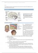
The folds of the brain are called
gyri and the grooves are called
sulci. Ventricles in the brain are
a communicating network of
cavities filled with cerebrospinal
fluid.
COVERING THE BRAIN
The brain has to be very
well protected and is therefore
surrounded by the skull (bone),
layers of various membranes (which
have blood vessels and nerves of
their own) and spinal fluid (which
cushions it).
These layers are perfectly fit
for the exact measurements of your
brain - ridges, and all. This isn’t very
good because, for example, if you hit
your head, your brain would slightly
change its position - as it changes
position, it could get scratched due to the ridges within the layers causing brain damage.
Explanation of layers:
► Skull - Rigid bone
► Dura mater – a thick layer
► Arachnoid membrane – a spongy layer. Blood (vessels) and spinal fluid flow through
here
► Pia mater – a delicate membrane that sits on the brain itself
The brain is stuck inside this limited space within the head.
► whenever it swells up, it pushes up against the protective layers. If important parts
of the brain are being pushed (squeezed), it hinders its performance, and you could
lose important bodily functions.
► When a blood vessel pops (causing a haemorrhage – bleeding in the protective
layer), the blood takes up space causing the brain to be squished. This causes the
brain to become desperate for space thereby pushing parts of itself (hindbrain) into
1
,Block 3 – Biological and cognitive psychology | Lecturer: Enid Schutte
a space near the bottom of the brain area (where the spine goes through) called foam
magnum.
THE ORGANISATION OF THE BRAIN
The brain’s three basic units:
Hindbrain – (essential function) regulates autonomic functions, coordinates movement,
maintains balance and equilibrium
Midbrain – processes auditory, and visual
information
Forebrain – processes sensory information,
responsible for thoughts, regulates motor
functions
HINDBRAIN
The hindbrain consists of:
Medulla Oblongata - Involuntary behaviours like breathing and circulation
Pons - Relay station between the brain and the spinal cord
(keeps asleep or awake)
Cerebellum - Coordinated movement, maintains balance
and posture.
Reticular formation (portions) - Arousal and consciousness
Reticular formation begins in the hindbrain and continues through to the
midbrain.
MIDBRAIN
Located above the pons.
Concerned with certain sensory processes such as locating where things are.
Inferior Colliculus – receives visual input
Superior Colliculus – receives auditory input
Substantia Nigra – produces dopamine (happy
transmitter, too much causes hallucination, too little
causes Parkinson’s disease).
Reticular Formation
► Runs through the hindbrain and the midbrain
► Contributes to the modulation of muscle reflexes,
breathing and perception of pain
2
,Block 3 – Biological and cognitive psychology | Lecturer: Enid Schutte
► Regulates sleep and wakefulness
► Ascending fibres within the reticular formation are essential to maintaining an alert
brain.
FOREBRAIN
The forebrain is covered by the cerebral cortex and consists of the:
Thalamus – initial processing of sensory information (except smell). Relay station for
sensory input
Hypothalamus – the pituitary gland, emotions, sleep, sexual activity, temperature
regulation, hunger, and thirst
Limbic system -
► Amygdala – emotions (e.g., fear), learning and emotional memory
► Hippocampus – controls and contains memory
Basal ganglia – movement. Planning of motor movement and aspects of memory and
emotional expression
Cerebrum -
► Contains the lobes. Lobes are areas bordered by major grooves and are defined by
their functions within the forebrain.
► Frontal lobe –
▷ Motor cortex – receives information from the spinal cord, cerebellum, and
basal ganglia; involved in voluntary movements. Initiates activity.
▷ Association areas – personality and higher-order thinking such as
organisation.
▷ Broca’s area – speech and controls expressive language
▷ Prefrontal executive function –
Dorsolateral, planning and
organising
▷ Takes the longest to develop.
► Parietal Lobes –
▷ Visual attention, integration of
different senses, locating the
position of objects in space, touch,
detection of movement and spatial
orientation
▷ Somatosensory cortex – mirrors the motor cortex, receives sensory
information, detection of movement
► Temporal Lobes –
▷ hearing and language function
▷ Wernicke’s area – responsible for understanding speech. Memory
acquisition. Categorization of objects. Visual associations.
► Occipital Lobes –
▷ Everything Vision
3
, Block 3 – Biological and cognitive psychology | Lecturer: Enid Schutte
▷ First to develop
What happens if the lobes are damaged or dysfunctional?
Frontal Lobe:
Paralysis – Loss of simple movement
Sequencing – inability to plan a sequence of complex movements needed to complete a
multi-stepped task
Loss of spontaneity in interacting with others
Perseveration – inability to focus on a task
Emotionally Labile – mood changes
Broca’s Aphasia – Inability to express language
Parietal Lobe:
Anterior region: numbness or impaired (what and where) sensation contralaterally
Bilaterally: Balint’s syndrome – visual attention and motor syndrome
Left hemisphere damage: right-left confusion, agraphia, visual agnosia (Gerstman’s
syndrome)
Right hemisphere damage: hemineglect, constructional apraxia, anosognosia (denial of
problems)
Apraxia: inability to perform tasks or movements when asked even though the quest was
understood
Temporal Lobe:
Left-Wernicke’s Aphasia: difficulty understanding words (tied with expression and
context)
Right-Prosopagnosia: difficulty in recognising faces
Associative Agnosia
Occipital Lobe:
Cortical blindness/ visual field cuts: defects in vision
Achromatopsia: difficulty identifying colours
Akinesthesia: inability to recognise/ interpret the movement of objects
Alexia and Agraphia: word blindness with writing impairments
Hallucinations and visual illusions
The Diencephalon of the forebrain is made of the thalamus, hypothalamus and epithalamus (which
creates brain and spinal fluid which washes away bad things).
LATERALISATION OF THE BRAIN
the brain is separated into the left and right hemispheres which are joined together by the
corpus callosum.
Lateralisation = certain functions are dominant in one or another hemisphere.
4


