100% satisfaction guarantee Immediately available after payment Both online and in PDF No strings attached
logo-home
SUMMARY chapter 1-10 The Immune system; Peter Parham (4th edition) R119,82   Add to cart

Summary

SUMMARY chapter 1-10 The Immune system; Peter Parham (4th edition)

 15 views  1 purchase
  • Course
  • Institution
  • Book

In this document, I have summarized the first 10 chapters of the immune system, written by Peter Parham. Not every paragraph is included (most of them I have covered). For Biomedical Sciences master students at the VU, this is also prior knowledge that has to be revised before starting the course '...

[Show more]
Preview 8 out of 75  pages

  • No
  • Chapter 1-10
  • August 29, 2024
  • 75
  • 2024/2025
  • Summary
avatar-seller
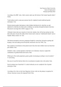
Chapter 1
Elements of the immune system and their roles in defence

1.4 The innate immune response causes inflammation at sites of infection

The innate immune response consists of two parts
1. Recognition that a pathogen is present
a. Involves soluble proteins and cell-surface receptors that bind either to the
pathogen and its products or to human cells and serum proteins that become
altered in the presence of the pathogen.
2. The recruitment of destructive effector mechanisms that kill and eliminate the pathogen.

Effector mechanisms
- Effector cells
- Complement (serum proteins)

When a pathogen is recognised by
cells and proteins, cytokines are send
out that interact with other cells to
trigger the innate immune response.
They induce the local dilation of blood
capillaries, that eventually leads to
increased blood plasma into the
damaged tissue, that contains
inflammatory cells (WBC).


1.5 the adaptive immune response adds to an ongoing innate immune response

Lymphocytes (WBC) are called while the innate response is slowing the spread of the infection.
While the effector mechanisms of the adaptive response are similar to those of the innate, the
important difference lies in the way in which lymphocytes recognize pathogens.


1

,recognising differences
- The receptors of the innate immunity comprise many different types that each recognize
features shared by groups of pathogens and are not specific for a particular pathogen.
- Lymphocytes (Adaptive) recognize pathogens by using cell-surface receptors of just one
molecular type.
o The proteins can be made in billions of different versions, each capable of binding
a different ligand. So it can be made specific for a particular pathogen
▪ The lymphocyte receptors are not encoded by conventional genes but by
genes that are cut, spliced, and modified during lymphocyte
development.

When the right lymphocytes have been chosen, they will proliferate and differentiate to produce
large numbers of effector cells specific for that pathogen.
Clonal selection: selecting the small subset of pathogen-specific lymphocytes for proliferation
Clonal expansion: selecting the small subset of pathogen-specific lymphocytes for differentiation

1.7 immune system cells with different functions all derive from hematopoietic stem
cells


Leukocytes, another term for all the white blood cells (WBC), derive from a common progenitor
called the pluripotent hematopoietic stem cell. From this stem cell, WBC, but also erythrocytes
and megakaryocytes (source of platelets) derive. An umbrella term for all these cells, plus their
precursor cells is hematopoietic cells.


These hematopoietic cells can do two things;
- Self-renewal: divide to give further hematopoietic stem cells
- Mature into three different cell lineages
1. Erythroid (RBC)
2. Myeloid (innate)
a. Granulocytes: have prominent cytoplasmic granules containing reactive
substances that kill microorganisms and enhance inflammation.
i. Neutrophil: specialized in the capture, engulfment and killing of
microorganism. It is a type of phagocytes (engulfing method).
Neutrophils are the most lethal of the phagocytes. They rapidly
mobilize to enter sites of infection and can work in anaerobic

2

, conditions! They are short lived and die at the site of infection,
forming pus
ii. Eosinophil: defends against helminth worms and other intestinal
parasites
iii. Basophil: helps the immune response to parasites, but little is
known of its contribution to immune defence.
b. Monocytes: circulate in the blood. They are big, have distinctive indented
nucleus and look all alike.
i. Macrophages: travel in the blood to tissues (where they mature) and
take up residence. It works, just like a neutrophil, by phagocytosis.
They have numerous vacuoles in their cytoplasm, often containing
engulfed material. They are also called the scavenger cells of the
body. Unlike neutrophils, they live long! They also provide warning
to other cells and orchestrate the local response to infection. They
secrete the cytokines that recruit neutrophils and other leukocytes
into the infected area.
ii. Dendritic cells: Have a distinctive star-shaped morphology. They
are similar to macrophages, but their unique function is to act as
cellular messengers that are sent to call up an adaptive immune
response when it is needed. If it is needed, they will leave the
infected tissue with a cargo of intact and degraded pathogens and
take it to lymphoid organs that specialize in making adaptive immune
responses
c. Mast cell: unknown precursor cell! Resident in all connective tissues. The
activation and degranulation of mast cells at sites of infection make major
contributions to inflammation.
3. Lymphoid (adaptive)
a. Natural killer cells: NK cells are important in the defence against viral
infections. They enter infected tissues, where they prevent the spread of
infection by killing virus-infected cells and secreting cytokines that impede
viral replication in infected cells.
b. T-cells:
i. Cytotoxic T cell: kill cells that are infected with viruses or with certain
bacteria that live inside human cells. (NK cells do the same, but
during the innate response).
ii. Helper T cell: secrete cytokines that help other cells of the immune
system become fully activated effector cells.
iii. Regulatory T cell: controls the activities of the cytotoxic and other
types of T cell, thereby preventing unnecessary tissue damage and
stopping the immune response once the pathogen has been
defeated.
c. B-cells:
i. Plasma cell: secrete immunoglobulins called antibodies that circulate
in the blood and can enter infected tissues. This type of immunity is
also called humoral immunity. The most important function of
antibodies is to facilitate the engulfment and destruction of
extracellular microorganisms and toxins by phagocytes. There are
two ways to reduce infection by antibodies;
1. Neutralization: binding tightly to a site on a pathogen so as to
inhibit pathogen growth, replication, or interaction with human
cells.


3

, 2. Opsonization: the coating of a bacterium with antibodies that
facilitates phagocytosis.

1.12 adaptive immunity is initiated in secondary lymphoid tissues

Dendritic cells with their pathogen cargo, travel to
the nearest lymph node by the lymphatics and
arrive in afferent vessels. In the lymph node you
have T-cell areas and B-cell areas, known as
lymphoid follicles. The dendritic cells settle in the
node and pathogens are filtered out by
macrophages. This prevents infectious organisms
from reaching the blood and provides a depot of
pathogenic material that can be used to activate
lymphocytes. The B cells are proliferating in the
germinal center to effector B cells, T cells into
effector T cells in the T cell area. When exiting the
node, via the efferent lymphatic vessel, the effector
B and T cells go to the infected tissues and work
with the innate immunity to subdue the infection.

1.13 the spleen provides adaptive immunity to blood infections

Some pathogens enter the bloodstream directly. For this, the spleen is the lymphoid organ that
serves as a filter for the blood. Purposes of this filtration are
1. To remove damaged or senescent red cells;
2. To sense other pathogens, to which the spleen works as a secondary lymphoid organ
against blood-borne pathogens.
The red pulp of the spleen is to monitor and
remove red blood cells, and the white pulp is
where WBC gateher to provide adaptive
immunity.


Chapter 2
Innate Immunity: the Immediate Response to Infection


Figure above → many barriers prevent pathogens from crossing epithelia and colonizing tissues.


4

,2.3 complement is a system of plasma proteins that mark pathogens for destruction

One of the first weapons to fire is a system of soluble proteins made
constitutively by the liver and present in the blood, lymph, and
extracellular fluids. They are known as the complement (system).
Complement coats the surface of bacteria and extracellular virus
particles and makes them more easily phagocytosed.

Especially bacteria that
The complement system consists of are enclosed in thick
mostly: polysaccharide capsules
- Proteolytic enzymes resist phagocytosis
- Proteases without complement!
They circulate in functionally inactive
forms known as zymogens. Infection
triggers complement activation, that results in proteases cleaving and
activating the next protease in the pathway. Enzymes are highly specific
for the complement it cleaves.

More than 30 proteins make up the complement system, but C3 is the most
important. Cleaving C3 leads to C3a and C3b, where C3b bounds to the The high-energy
pathogen’s surface. This complement fixation is essential because it makes the thioester bond within
bacteria more eligible for phagocytosis destruction. In addition, it organizes the the glycoprotein is
formation of protein complexes that damage the pathogen’s membrane. The C3a what makes C3 so
fragment also contributes, by acting as a chemoattractant to recruit effector cells, unique!
including phagocytes, from the blood to the site of infection.

There are 3 pathways that all lead to C3 activation:


2.4 At the start of an infection, complement activation proceeds by the alternative
pathway
This is used especially against bacterial infection.

5

,1. At a slow rate, without being cleaved, the thioester bond that was in its inactive form,
changes its conformation and exposed the bond. In the aqueous blood the bond
becomes active and quickly makes a covalent bond that attaches C3 to another
molecule (usually H2O) called iC3/C3(H2O).
2. An infectious environment increases the rate at which C3 is hydrolyzed to give iC3. iC3
binds to the inactive complement factor B, making factor B susceptible to cleavage by
the protease factor D.
3. This reaction produces Ba (gets released) and Bb (remains bound to iC3 and has
protease activity).
a. The new complex iC3Bb is a protease that specifically and efficiently cleaves C3
into the C3a and C3b fragments, with exposure of the thioester
bond that is in C3b. Proteases that cleave
4. This way, with large number of C3 molecules being cleaved and and activate C3 are
activated, some C3b fragments become covalently attached to amino called C3 convertases.
and hydroxyl groups of the pathogen’s outer surface.
5. Pathogen-bound C3 binds factor B and facilitates the cleavage of factor B by factor D.
this reaction leads to the release of Ba and the formation of a C3bBb complex on the
microbial surface
a. C3bBb is a potent C3 convertase, called the alternative C3 convertase, which
works right at the surface of the pathogen. It binds C3 and cleaves it into C3a
and C3b with activation of the thioester bond. Because C3bBb is fixed to the
pathogen’s surface and doesn’t diffuse away, the C3b cleaved fragments become
quicker fixed to the pathogen.
6. In the end, it is a positive-feedback process, in which the C3b product of the reaction
can assemble more enzyme, is one of progressive amplification of C3 cleavage.


6

,2.5 Regulatory proteins determine the extent and site of C3b deposition

Increases the reaction: The plasma protein properdin
(factor P) increases the speed and power of
complement activation by binding to the C3 convertase
C3bBb on microbial surfaces and preventing its
degradation by proteases.

Inhibits the reaction: Plasma protein factor H, also
binds to C3b and facilitates its further cleavage to a
form called iC3b by the plasma serine protease factor I.
fragment iC3b cannot assemble a C3 convertase, so
the combined action of factors H and I is to decrease
the number of C3 convertase molecules on the
pathogen surface.

Inhibiting on human cell surface:
Decay-accelerating factor (DAF) binds to the C3b
component of the alternative C3 convertase, causing
its dissociation and inactivation.

Membrane cofactor protein (MCP) also has this
function, but the binding of MCP to C3b makes it
susceptible to cleavage and inactivation by factor I.
MCP is similar to factor H

Macrophages in the liver
2.6 Phagocytosis by macrophages provides a first line of cellular are known as Kupffer cells
defence against invading microorganisms. --
The coating of a pathogen
with a protein that
facilitates phagocytosis is
The process of phagocytosis by macrophages is made more efficient by called opsonization
receptors on the macrophage surface.
- Complement receptor 1 (CR1): binds to
C3b fragments that have been deposited at
high density on the surface of a pathogen
through activation of the alternative pathway
of complement.
- CR3 and CR4: bind to iC3b fragments on
microbial surfaces. iC3b facilitates
phagocytosis and pathogen destruction by
serving as the ligand for CR3 and CR4.


Together these three receptors work more effectively than they work on their own!


7

, 2.7 The terminal complement proteins lyse pathogens by forming membrane pores


There are next to the C3 complement,
five additional complement
components. These components all
belong to a cascade that eventually
form into the membrane-attack
complex. This makes holes in the
membranes of bacterial pathogens and
eukaryotic cells. They are listed here
right.


On human cells
On human cells it is regulated by soluble and surface-associated proteins.
- The soluble proteins S protein, clusterin,
and factor J prevent the soluble complex
of C5b with C6 and C7 from associating
with cell membranes.
- At the human surface, the proteins
homologous restriction factor (HRF) and
CD59 (also called protectin) prevent the
recruitment of C9 by the complex of C5b,
C6, C7 and C8.


2.8 small peptides released during complement activation induce local inflammation

The C3a or C5a fragments of the complements increase
inflammation at the site of complement activation through
binding to receptors on several cell types.

In some circumstances, the C3a and C5a fragments induce
anaphylactic shock, an acute inflammatory reaction that
occurs simultaneously in tissues throughout the body. They
are referred to as anaphylatoxins. C5a is more stable.
- The anaphylatoxins induce the contraction of smooth
muscle and the degranulation of mast cells and
basophils, with the consequent release of histamine
and other vasoactive substances that increase
capillary permeability.
- They also have direct vasoactive effects on local
blood vessels, increasing blood flow and vascular
permeability.
➔ These changes make it easier for plasma proteins
and cells to pass out of the blood into the site of an
infection

8

The benefits of buying summaries with Stuvia:

Guaranteed quality through customer reviews

Guaranteed quality through customer reviews

Stuvia customers have reviewed more than 700,000 summaries. This how you know that you are buying the best documents.

Quick and easy check-out

Quick and easy check-out

You can quickly pay through EFT, credit card or Stuvia-credit for the summaries. There is no membership needed.

Focus on what matters

Focus on what matters

Your fellow students write the study notes themselves, which is why the documents are always reliable and up-to-date. This ensures you quickly get to the core!

Frequently asked questions

What do I get when I buy this document?

You get a PDF, available immediately after your purchase. The purchased document is accessible anytime, anywhere and indefinitely through your profile.

Satisfaction guarantee: how does it work?

Our satisfaction guarantee ensures that you always find a study document that suits you well. You fill out a form, and our customer service team takes care of the rest.

Who am I buying this summary from?

Stuvia is a marketplace, so you are not buying this document from us, but from seller jb3802. Stuvia facilitates payment to the seller.

Will I be stuck with a subscription?

No, you only buy this summary for R119,82. You're not tied to anything after your purchase.

Can Stuvia be trusted?

4.6 stars on Google & Trustpilot (+1000 reviews)

73091 documents were sold in the last 30 days

Founded in 2010, the go-to place to buy summaries for 14 years now

Start selling
R119,82  1x  sold
  • (0)
  Buy now